Smokes your problems, coughs fresh air.

Tag: nutrition (Page 1 of 2)

Metabolism & Nutrition: learning objectives

Lectures

Energy metabolism

  • “Explain the Harris-Benedict principle.”

    Mean energy requirements are ~2000–2400 kcal/day, slightly less for ♀♀ than for ♂♂. Basal Metabolic Rate (BMR) can be estimated using the Harris-Benedict equation:

    BMR = 10 x weight (kg) + 6.25 x height (cm) – 5 x age (yrs) + 5 if ♂
    BMR = 10 x weight (kg) + 6.25 x height (cm) – 5 x age (yrs) – 161 if ♀

    According to this equation my BMR = 10 x 75 + 6.25 x 182 – 5 x 33 + 5 =
    750 + 1137.5 – 165 + 5 = 1728 kcal/day.

    Multiply BMR with a number between 1.2 and 1.9, depending on daily activity level.

  • “Name the energy-content and storage of three energy-carriers.”
    Macronutrient class E (kcal/g)
    Carbs 4
    Proteins 4
    Fats 9
    Alcohol 7
  • “Outline the interaction between energy supplying metabolic pathways.”

    Blood [glucose] has to remain ≈ 5mM. Lower levels will trigger the release of glucagon

    ≤ 1 day of fasting:
    glucagon (pancreatic alpha cells) → glycogenolysis (in muscle and liver tissue).
    glucose (liver) → systemic release (through glucose-6-phosphate)
    >1 day of fasting:
    triglyceride store,
    gluconeogenesis by muscle catabolization → ketons
    blood [glucose] < 5mM
    glucagon ↑ (pancreas)
    blood [glucose] > 5M
  • “Describe the regulation of the glycolysis/gluconeogenesis switch.”

    Muscle tissue, at rest: Glycolysis (hexokinase) inhibited by its product [Glucose 6-Phosphate] increasing; pyruvate kinase inhibited by increasing [ATP]/[AMP] ratio.

    Glucose → F-6P → F-1,2-BP + F-2,6-BP
    Fructose 2,6-biphosphate activates PFK, thus upregulating glycolysis

    Low [ATP]/[AMP]: PFK ↑. PFK catalyses F6-P → F1,6-BP + ATP
    Liver: no negative feedback of [Glucose 6-Phosphate] on hexokinase [because no hexokinase].

    Muscle tissue, during exercise:

    Switch from glycolysis to gluconeogenesis occurs fructose-2,6-biphosphate

  • “Name the most important steps in the Krebs cycle.”

    First, glycolysis occurs in the cytosol:

    • Pyruvate → Acetyl-Coenzyme A (CoA) (1CO₂ + 2e¯)

    Krebs cycle / citric acid cycle happens within the mitochondrial matrix:

    1. C₂ (Acetyl-CoA) + C₄ (oxaloacetate)→ C₆citrate (2CO₂ + 8e¯)
    2. C₆ → C₅ + NADH + CO₂
    3. C₅ → C₄ + NADH + CO₂
    4. C₄ → C₄ (oxaloacetate) + NADH + FADH₂ + ATP
  • from Acetyl-CoA → 2CO₂ + 3NADH + FADH₂ + ATP:
    8e¯ used to form 3NADH + FADH₂;
    2C + 4O used to form 2CO₂

  • “Describe the different components of oxidative phosphorylation.”
    1. A physical link to the citric acid cycle;
    2. four complexes (I-IV); and
    3. three proton pumps.

    Inputs: 8 e¯ (carried by NADH⁺ and FADH₂ from the citric acid cycle) + 2O₂
    Outputs: ATP + 4H₂O

  • “List the locations of the different metabolic pathways in the cell.”
    glycolysis cytosol
    Krebs cycle mitochondrial matrix
    oxidative phosphorylation mitochondria

Fat metabolism

  • “Describe the chemical structure of saturated and unsaturated fatty acids, of phospholipids, of triglycerides and of cholesterol.”
    Saturated fatty acids
    No double bonds between C atoms; all carbons are ‘saturated’.
    Unsaturated fatty acids
    One or more double bonds between (some) C atoms.
    Phospholipids
    Hydrophobic tail and hydrophilic head.
    Triglycerides

    chemically inert, energy-dense
    Cholesterol
    Many apolar structures (only 1 OH group), making it very hydrophobic.
  • “Summarize how fatty acids are lysed, absorbed by intestinal cells and transported through the body.”

    Lumen: triglyceride + lipase → FFAs + monoacylglycerol
    Mucosal cells: reassembly of triglyceride → incorporated in chylomicrons
    Lymph system

  • “List the characteristics, structure, and physiological role of 5 different lipoprotein particles.”
    Lipoprotein particle Diameter (nm) Composition Physiological role
    Protein TG C CE PL
    Chylomicrons 500 2% 85% 1% 3% 9% Transport of fat to tissues
    VLDL 43 10% 50% 7% 13% 20% Carry newly synthesized triglycerides from the liver to adipose tissue
    IDL 27 18% 26% 12% 22% 22% Not usually detectable in the blood during fasting
    LDL 26–27 25% 10% 8% 37% 20% Transport of fatty acids and cholesterol from the liver to other tissues
    HDL 9.5, 6.5 55% 4% 2% 15% 24% Collect fat molecules from cells/tissues, and take it back to the liver

  • “Explain how the body mobilizes fat during fasting and how this is regulated.”

    Glucogen triggers
    Adipose cells: triacylglycerol (=triglyceride) → FFAs + glycerol
    Liver cells: FFAs and cholesterol built into VLDL particles; glycerol → pyruvate (glycolysis), glycerol → glucose (gluconeogenesis)
    Other tissues: FFAs oxidized → Acetyl CoA → citric acid cycle (→ CO₂ + H₂O)

  • “List the advantages of energy storage in the form of triglycerides.”

    Triglycerides are very energy-dense: 9 kcal/g; hydrophobic; chemically inert; no important functional role; hydrophobic: the energy-equivalent of 12kg of fat in glycogen would require 100kg of water.

  • “Point out the relationship between unsaturated fats, trans-fats and cardiovascular disease.”

    unsaturated fats: VLDL↓ & HDL↑ => P(CVD)↓
    trans-fats: LDL↑ & HDL↓ => P(CVD)↑
    too much saturated fat: P(CVD)↑, P((colonic) cancer)↑, P(type II diabetes)↑
    insufficient unsaturated fat: P(type II diabetes)↑

  • “Explain the biological role of cholesterol.”

    Cholesterol is essential for synthesizing cell membranes and for metabolites, including:

    • steroid hormones (androgens, estrogens, progestins, glucocorticoids, and mineralocorticoids); and
    • bile acids (detergent function for lipid absorption and biliary secretion, signaling function).
  • “Describe how cholesterol synthesis is regulated.”

    The molecular regulation of cholesterol synthesis follows a general feedback regulation system, wherein, when a precursor is converted into a product by an enzyme, decreasing [product] upregulates the enzyme (through a sensor) and increasing [product] downregulates the enzyme (through another sensor). The cholesterol sensor is SCAP (SREBP cleavage activating protein), which, in the presence of sufficient cholesterol, binds to an ER protein called Insig. SCAP escorts SREBP (sterol regulatory element binding protein) from the ER to the Golgi apparatus in the absense of cholesterol.

  • “Define the differences between LDL and HDL.”

    LDL transports fatty acids and cholesterol from the liver to other tissues.
    HDL collects fat molecules from misc. tissues for transport back to the liver.

    [Learn diagram of damage by LDL and ‘repair’ by HDL.]

  • “Describe the synthesis and metabolism of HDL.”

    HDL is synthesized in the hepatocytes of the liver and in the enterocytes of the intestine. In these cells (almost exclusively), the Apolipoprotein A1 (APOA-I) gene is expressed, which is a major protein component of HDL. Cholesterol efflux to APOA-I is mediated by the ubiquitous ABCA1 transporter.

    [And what about metabolism?]

  • “Reproduce the cholesterol absorption route.”

    exogenous C → chylomicrons (intestine) → VLDL → LDL → perihpial tissues

    [Study slides]

  • “Describe the complete reverse cholesterol transport route and the characteristics of the TICE route.”

    Reverse cholesterol transport

    Cholesterol in macrophages in periphial tissues is transported as HDL to the liver, which excretes it as bile into the intestine.

    Transintestinal cholesterol transport (TICE)

    TICE is a direct route of HDL from the intestine to the periphial tissues. It is an active payway facilitating the direct excretion of cholesterol from the enterocyte, likely mediated by apoB-containing lipoproteins.

  • “Indicate why HDL particles are healthy.”

    HDL

    • promotes cholesterol efflux,
    • promotes reverse cholesterol transport,
    • inhibits LDL oxidation,
    • inhibits endothelial inflammation,
    • promotes endothelial NO production,
    • promotes prostacyclin availability, and
    • inhibits platelet aggregation.

Carbohydrate metabolism

  • “List the different types of carbohydrates.”
    • Monosaccharides: glucose, fructose, galactose;
    • disaccharides: saccarose, lactose, maltose, isomaltose;
    • polysaccarides: α-glucosides (starch, glucogen), β-glucosides (cellulose).
  • “Summarize the digestion and transport routes of carbohydrates.”

    Only monosaccharides are absorbed in the small intestine. Poly- and disaccarides first have to be split:
    Starch → glucose;
    lactose → galactose + glucose;
    saccharose → fructose.

    Fructose absorbed by GLUT5 transporter;
    glucose/galactose by SGLT1.

  • “Describe why and how blood glucose values are regulated.”

    Insulin stimulates anabolic pathways (storage, synthesis, etc.), so that proteins are synthesized.
    Glucagon stimulates catabolic pathways (breakdown, energy production), so that proteins are broken down.

  • “Explain the difference between glucokinase and hexokinase.”

    Glucokinase (in pancreas, liver and brain cells) has a much lower KM (5.5 mmol/l) than hexokinase (in most other cells) so that insulin secretion of pancreatic B-cells can be positively correlated to blood plasma [glucose].

  • “Describe the glycogen metabolism.”

    Glycogen → Glucose 1-phosphate → Glucose 6-phosphate

    Fasting (low glucose): glucagen released by α-cells in the pancreas. Glucagen stimulates the conversion of glycogen to glucose, as does adrenaline.

    Eating (high glucose): insulin produced by β-cells in the pancreas prompts muscle and liver cells to convert glucose to glycogen.

  • “Describe what it means to be lactose intolerant.”

    Lactose-intolerent people have no lactase production or produce inactive lactase.

Protein and amino acid metabolism

  • “Describe how proteins are degraded into amino acids.”

    Dietary protein degradation

    In the mouth: denaturation starts;
    in the stomach: HCl and pepsine further break and hydrolize the proteins into polypeptides;
    in the small intestine: proteases (e.g. aminopeptidase) break the polypeptides further down, into amino acids and dipeptides.

    Intracellular protein degradation

    Ubiquitinated protein → proteasome → peptide fragments + ubiquitin;
    proteolysis: peptide framments → amino acids

  • “Indicate the difference between essential and non-essential amino acids.”

    Essential amino acids cannot be synthesized by the body and have to be included in dietery sources.

  • “Indicate the location of protein degradation.”

    In the mouth, the stomach and the small intestine. Also, intracellularly.

  • “Describe the glucose-alanine cycle.”

    In the muscle: glucose →(glycolysis)→ pyruvate; branched-chain amino acids → NH₄⁺; pyruvate + NH₄⁺ → alanine
    Alanine is transported to the liver, where it can be used in gluconeogenesis (alanine → pyruvate → glucose) or converted to urea (alanine → glutamate → NH₄⁺ → urea).

  • “Describe the urea cycle.”

    Amino acid degradation takes place mostly in the liver: alpha-amino acid → glutamate → ammonium (NH₄+) → Urea (with NH₂ groep). (Urea cycle takse two NH₄+ and Fumerate [Gluconeogenesis lecture]. Fumerate is another of the amino acid breakdown products (not in the urea cycle).)

  • “Describe how amino acids are used as an energy source.”

    Gluconeogenesis: CO₂ + NH₄⁺ → urea cycle. In one of the next steps, aspartate (one of the products of an alpha-amino acid + oxaloacetate) enters the cycle: citrulline + aspartate → arginino-succinate → fumarate + arginine.
    fumarate → malate → oxaloacetate → glucose + aspartate.

  • “Describe situations in which there is an abnormal protein requirement.”
    • Infancy
    • Pregnancy
    • Malabsorbtion of amino acids
    • Metabolic disease (e.g. PKU/-phe)
    • Protein sensitivity / bovine protein intolerance
    • Diseaese/trauma/wounds

Metabolic regulation

  • “List the transcription factors and associated ligands which are important for the regulation of the energy metabolism.”

    LXR (liver X receptor) is activated by cholesterol. In the liver, LXR activates ABCG5/8, inceasing the release of Chol+BA by hepatocytes. This, in turn, lowers blood cholesterol, as well as atherosclerosis, but also raises triglyceride production. LXR is thus a promising target for treating atherosclerosis, with the unintended side-effect of weight-gain. Indeed, a synthetic LXR ligand (GW3965) has been found to inhibit the development of atherosclerosis in LDLR and apoE knockout mice by S.B. Joseph et al. (PNAS 2002;99:7604-7609). And, indeed, the treated mice gained weight as well.
    LXR also stimulates the absorption of FFA by the hepatocytes.

    FXR (farnesoid X receptor) is activated by bile acids. FXR then activates MDR2 and BSEP transporters (which facilitate the release of PC and BA, respectively). FXR lowers cholestasis, the incidence of chol. gallstones, and the production of triglycerides; as a target for weight-loss, the unintended side-effect will be atherosclerosis.

    PPAR (Peroxisome Proliferator-Activated Receptor) family of receptors bind to fatty acids. PPARγ is implicated in energy storage. PPARδ and PPARα are implicated in energy burning.

  • “Explain how a nuclear hormone receptor works.”

    Ligand-mediated activation: nuclear receptor (NR) binds to HRE (Hormone Receptor Element), a stretch of DNA to which also an RXR nuclear receptor is bound. This complex is bound by a HDAC co-repressor complex, actively repressing the HRE. When a ligand binds to the NR, the HDAC co-repressor complex is displaced by the HAT co-activator complex, which causes acetylization of a DNA region adjacent to the HRE. (HDAC = Histone Deacetylase; HAT = Histone Acetyl Transferase.) Acetylization activates the genes.

  • “List the characteristics of the metabolic syndrome.”

    Obesity, high blood sugar, high LDL, high blood TG, high blood pressure.

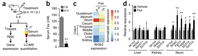
  • “Name drugs that act through the activation of nuclear hormone receptors.”
    • Fex selectively activates intestinal FXR.
    • Avandia activates PPARγ, causing the burning of FFAs.
  • “List processes that take place during the enterohepatic cycle of bile salts.”

    FXR is expressed in the liver, which releases bile salts. Bile, produced by hepatocytes (liver cells) ends up in bile ducts, aided by 3 ABC transporters of relevance: ABCG5/8 transporters (for cholesterol), MDR2 transporters (for PC=phosphatidylcholine), and BSEP transporters (for BA). Bile is made up of 4% cholesterol, 24% PC and 72% BA. PC helps to neutralize BA so that it doesn’t damage cell membranes.

    BA reenters the cell with the aid of a transporter called NTCP. Cholesterol also enters the hepatocytes (liver cells) (LDL through LDLR, HDL through SR-B1).

    FXR activation triggers the release of bile from the hepatocyte. In enterocytes (in the illeum, the final section of the small intestine), FXR downregulates the uptake of BA by ASBT transporters and stimulates the release of BA by OSTa/b. FXR is also responsible for the release of bile salts from the illeum into the capillaries. Bile salts are also released into the blood stream by the gallbladder, which constricts under the influence of CCK.

  • “Decribe the regulation of fat metabolism in the liver.”

    Triglyceride (TG) from the diet or adipose tissue is hydrolysed (lipolysis) into free fatty acids (FFA), which enters the hepatocyte through the CD36 transporter. FFA in the hepatocyte can be converted into TG and stored in a complex with cholesterol, or TG+chol. can leave the hepacotye through MTTP as VLDL. Alternatively, FFAs can be used in the FAO pathway for the energy necessary to produce ketone bodies during the production of Acetyl CoA.

    LXR activates lipogenesis (FFA synthesis from sugars) and increases FFA uptake from the blood, causing more TG production, which is dependent on [FFA]. ABCG5/8 transporters are also activated, increasing the release of Cholestorol and BA into the bile ducts. FXR, however, inhibits LXR and also directly inhibits lipogenesis.

  • “Describe the transcriptional regulation of different muscle fiber types.”

    PPARδ function remained unknown for a long time. It didn’t help that knockout mice died while still in the embryoic phase. The solution was to construct mice (VP16-PPARδ mice) in which PPARδ is only locally overexpressed, in muscle. VP16-PPARδ red muscle mass increased relative to controls.

    Transgenic mice with increased PPARδ in their muscles were true “marathon mice”. These mice are also overweight-resistent, which makes PPARδ a promesing anti-obesity drug-target. It as already used as doping by cyclists.

    PPARδ expression in muscle tissue → fast twitch,
    PPARδ suppression in muscle tissue → slow twitch.

  • [Study Slide 49 & 50.]

Integration of metabolism

  • “List different satiation signals.”
    • GLP-1, secreted by L cells in the small intestine, signals satiety in the brain; as does
    • CCK, which is also released by the small intestine.
    • Leptin is an adipokinese, a signal molecule from fat tissue.

    GLP-1 also causes the release of insulin by the pancreas.
    CCK also causes the constriction of the gall bladder.

  • “Indicate how leptin regulates the energy balance.”

    Normally, a decrease in fat cell mass → decrease in leptin expression → decreasing leptin action in hypothalamus → increase in food intake, while an increase in fat cell mass → increase in leptin expression → increasing leptin action in hypothalamus → decrease in food intake.

  • “Indicate the consequences of leptin resistence and deficiency.”

    Leptin-defficient (ob-/ob-) mice and men never stop eating.

  • “Indicate the consequences of excessive calorie intake.”

    Metabolic syndrome occurs when an excess of triacylglycerols can no longer be stored by the adipose tissue and is stored as lipid drops in other tissues. Insuline resistance results from energy stress (overnutrition and inactivity). Stress-induced serine kinases (which increase through the effects of mitochondrial overload and the increase of DAG and Ceramide) make GLUT4 less easily activated by the insuline receptor in the plasma membrane.

    Blood glucose rises with insulin resistence, which makes the pancreatic beta-cells work harder to produce more insuline, until, finally, the pancreatic cells give in and Type II diabetes turns into Type I diabetes. (During pregnancy, some measure insulin resistence is required. Maybe a contributing factor in insulin resistance is increased estrogen production by adipose tissues?)

  • “Describe the changes in energy metabolism during fasting.”

    The liver mobilizes the glycogen store: Glycogen → G1-P → G6-P → Glucose. (In the muscles and brain, glycolysis produces pyruvate from G6-P. Pyruvate is burned into CO₂ and H₂O in the brain and in aerobically exercised muscle, and into lactate in anaerobically exercised muscle.) However, this is only enough for 1 day.

    Gluconeogenesis uses amino acids to produce new glucose. Burning too much protein for energy is dangerous, though. The second priority is thus to maintain proteins. This is to some extent solved by ketone bodies [Fig 27.12/13], which can be formed from free fatty acids. Ketone bodies are hydrophilic and can pass the blood-brain barrier. Red blood cells still require glucose which is why the blood [glucose] has to remain > 2.2 mM.

Tutorials

Common objectives for all articles:

  • “List the most important conclusions from the articles treated.”
  • “Understand the principles upon which the articles are based.”

Diet

  • “Explain why specific metabolite concentrations have been measured in the study.”

    Blood [creatine] as an indicator of renal health;
    Blood [C-reactive protein (CRP)] as an inflammation marker;
    Urine [ketone] as an indicator of a glucose shortage, thus the burning of fat.

  • “Describe which metabolic adjustments take place in reaction to a low-carb or a low-fat diet.”

    In both studies,
    down: body weight, fat mass, triglyceride, CRP, 10-year Framingham CHD Risk Score;
    up: HDL,
    but for the people in the low-fat group everything goes up/down further.

  • “Formulate a follow-up study to the diet study.”

    Is het “laag-vet dieet” in deze studie wel een laag-vet diëet? Het zit nog steeds boven de aanbevelingen van het voedingscentrum.

    Measure BMR to check if low-carb increases resting energy expenditure and total energy expenditure.

Cardiovascular disease

  • “Describe where and how TMAO is produced.”

    phosphatidylcholine (PC) / choline → TMA → TMAO
    TMA (gasseous) is produced from PC/choline by intestinal microflora.
    TMA is converted to TMAO by FMO3 enzyme.

  • “Describe the possible mechanism how phosphatidylcholine influences the formation of artherosclerosis.”

    “Aortic macrophage content and scavenger receptor surface within aortic lesions were markedly increased in mice on the high-choline diet” (Wang et al. 2011).

  • “Point out possibilities of dietary interventions to lower the formation of TMAO.”

    Eat less red meat and other sources of choline.

Metabolic regulation

  • “Explain the difference between brown and white adipose tissue and their functions.”

    There is higher energy expenditure in brown adipose tissue (BAT), which is caused by the greater amount of mitochondria than in white adipose tissue (WAT).

  • “Point out the role of UCP1 in adipose tissue.”

    UCP1, which is expressed in BAT mitochondria, deflects the energy in the proton gradient to the production of warmth instead of ATP.

  • “Describe the effect of beta-adrenergic receptor (βAR) activation on fat cells.”

    The authors report increased UCP1 expression in adipose tissue. This might (partially) have been induced by increased beta adrenergic receptor (βAR) expression in WAT, through activation by catacholamine, which also explains the increase in FFAs and the decrease in triglycerides.

  • “Explain what the respiratory exchange ratio (RER) tells about energy usage and how you can measure this.”

    RER, which is the ratio VCO₂/VO₂, provides information on the active metabolic pathway. RER = 0.7 during the oxydation of carbohydrates and RER = 1.0 during the oxydation of fatty acids. RER ≤ 1.0. During the night, when most humans are asleep, we burn more fat than carbohydrates. For mice, which are nocturnal, the opposite is true.

    These values would have been supplied by the apparatus with which they performed energy expenditure measurements: a Comprehensive Lab Animal Monitoring System (Columbus Instruments).

  • “Explain what insulin and glucose tests are and what insight they provide into glucose homeostasis.”

    Hyperinsulinemic euglycemic clamp study: Mice are infused with glucose and the insuline response is measured. […]

Food and epigenetics

  • “List 3 basic epigenetic mechanisms.”
    1. DNA methylation,
    2. histone modification, and
    3. non-coding RNA.
  • “Eplain how DNA methylation can affect gene expression.”

    DNA methylation—the addition of a methyl group to cytosine or guanine nucleotides—affects the readability of the methylized DNA.

  • “List human examples of nutrient-epigenetic interactions (historic or geographic).”
    • Dutch Famine: children of mothers who suffered malnutrition during pregnancy showed an increase change of developing a number of metabolic diseases.
    • Barker Study in Wales: lower birth weight increased the chance of mortality through heart disease.
    • Gambian women: higher methylation of 6 metastable epialleles (MEs) in children conceived during (the meager) rainy season.
  • “Discuss the experimental planning of a diet/epidemiological study.”

    […]

Nutrition and microbiota

  • “List the effects of a vegetarian and an animal-based diet on the microbiome and its fermentation products.”

    “The animal-based diet increased the abundance of bile-tolerant microorganisms (Alistipes, Bilophila and Bacteroides) and decreased the levels of Firmicutes that metabolize dietary plant polysaccha-
    rides (Roseburia, Eubacterium rectale and Ruminococcus bromii). Microbial activity mirrored differences between herbivorous and carnivorous mammals2, reflecting trade-offs between carbohydrate and protein fermentation.” (David et al. 2014)

  • “Describe the relationship between the microbiome and obesitas.”

    Obesity of mice is partly determined by gut microbiota; if these microbiota are transplanted, the recipient mice will also become obese (Ley et al. 2006): “the relative proportion of Bacteroidetes [relative to Firmicutes] is decreased in obese people by comparison with lean people, and […] this proportion increases with weight loss on two types of low-calorie diet.” David et al. (2014) observed a reduction in weight for subjects that switched to an animal-based diet.

    More fermentation → more FA extracted from food.

  • “Indicate how the microbiome contributes to the development of children in relation to disease and what the role of nutrition is in this process.”

    […]

Nutrition and microbiata learning objectives from slides
  • “For four types of dietary fiber, list the effect on the microbiome, and on FA/butyrate production.”
    1. Inuline;
    2. resistant starch;
    3. cellulose;
    4. Gos/Fos (oligosaccharides).
  • “[…]”

Eatmeter

  • “Calculate the daily energy requirement.” \(
    BMR_♂(w=75, h=182, a=33) = C_♂ + W_♂w + H_♂h – A_♂a
    \)

    Depending on physical activity level, multiply BMR with a value between 1.2 and 1.9.

  • “List factors which influence the basal metabolic rate.”

    Age; gender; physical activity level.

  • “List the physiological function of dietary fibers.”
    • stommach filling;
    • bulk forming and nutrient dilution causing slower absorption in small intestine and less glucose spiking, while food uptake remains efficient;
    • fiber-nutrient interaction;
    • longer transit time;
    • a food source for microbiota.
  • “Indicate by which pathways the microbiome can influence the energy metabolism.”

    [See my notes on the Eatmeter tutorial.]

  • “List the fat-soluble vitamins and water-soluble vitamins, and explain the difference in terms of absorption, transport and storage of these vitamins.”

    Fat-soluble: A, D, E, K.
    Water-soluble: B, C.

    Absorption of fat-soluble vitamins goes up with increasing dietary fat uptake.

Caroric restriction

  • “Define ‘ageing’ and interpret the term in an evolutionary context.”

    Senescence: declining function with advancing age, among which decreasing fertility, and increasing mortality.
    Ageing is not universal. Hydra, for example, do not age. Hydra reproduce asexually, which may hint at the absense of a disposable soma. Because Hydra genes do not invest in genetic variation, reproduction may confer no advantage over simply staying alive.

  • “Explain ultimate and proximate ageing theories, and give examples.”

    Ultimate ageing theories explain why evolution has produced organisms that age, in terms of phylogeny and functional adaptation; while proximate theories explain how organisms age, in terms of ontogeny and causative mechanisms.

    The distinction between proximate and ultimate ageing theories is meaningless. Most ageing theories attempt to explain both proximate and ultimate questions. Evolutionary ecologists have to be concerned with all 4 of Tinbergen’s questions.

    A recurring concept in ultimate ageing theories is that wild animals do not often live to an age at which senescence comes into play, which severely limits the ability of natural selection to act on traits which would inhibit senescence.

    1. Mutation accumulation theory: senescence can persist in a population simply because natural selection doesn’t get a chance to come into play due
      to the low likelihood of individuals surviving long enough to reap a fitness benefit from decreased
      senescence. Then, old-age is caused by late-acting deleterious mutations which had no influence on reproduction (Medawar, 1952).
    2. Antogonistic pleiotropy: some late-acting deleterious genes may offer a benefit early in life, thus being favored by selection, even if selection forces are still active in later life (Williams, 1957). The slides say that the theory considers “no selection pressure on detrimental effects in postreproductive lifespan,” but this is false; it is definitely a theory of trade-offs between alleles that give an advantage during reproductive life and alleles that give an advantage during late life.
    3. Disposable soma: life-history trade-off between growth/reproduction and somatic maintenance/repair (Kirkwood, 1977).
    4. Programmed death / phenoptosis / acute senescence: a possible example are Pacific Salmon, which spawn immediately after spawning. Although this is normally assigned to exhaustian, Sapolsky (2004) points out that if a Salmon’s adrenal glands are remove right after spawning, it will live for quite a bit longer. Similar observations have been made in some species of Australian marsupial mice.
  • “Define the term ‘calorie restriction’ (CR).”

    CR is a 10–30% reduction in calories, below what would be eaten when feeding ad libitum.

  • “Apply principles outlined in ageing theories to the topic of caloric restriction.”
  • “Explain major differences between small lab animals and primates, which may lead to different responses to CR.”

    Small rodents are r-strategists, many offspring, short weaning-times, little energy invested per individual, short life-spans.
    Primates are K-strategists: few offspring, long weaning-time, lots of inergy invested per individual offspring, long life-spans.

    Compared to rodents, somatic maintenance and repair in primates is subject to much stronger and sustained selection forces, because parents have to grow old enough to take care of their young. This produces very different outcomes when considering disposable soma models. Additionally, grand-parental care can play a role, so that kin-selection must also be considered.

  • “Explain major considerations in the setup of calorie restriction studies in primates, especially regarding the measurement and implementation of CR.”

    […]

  • “Explain different setup and observations in the NIA and UW CR studies, and outline possible explanations.”
    NIA UW
    Study onset 1987
    Control group diet rationed ad libitum
    Supplementation control and CR CR only
    Age-related mortality no reduction sign. reduction
    Metablic health / overall function +
    Immune response improved in young-onset monkeys
  • “Explain the importance of control groups in CR studies.”

    Ehm, where else are you going to compare the CR treated subjects with?

  • “Name further factors, in addition to CR, that affect life and health span.”

    Genetics. Wild-caught mice showed no increase in life-span due to CR. Also worth mentioning that the NIA population was more genetically diverse.

Metabolism & Nutrition – Tutorial 3: Nutrition and epigenetics

The third tutorial for the RuG Metabolism & Nutrition course, was introduced and moderated by Torsten Plösch, who specializes in the epigenetic effects of nutrition during pregnancy.

The epigenetic effects of being conceived during the Dutch Famine

During his introduction, Torsten discussed the Dutch Hunger Winter 1944/1945 as a ‘natural’ experiment with which the effects of undernutrition can be studied. This Dutch Famine was a period of 4 months during which the average caloric intake of many adults in urban areas was only about 800-1000 kcal/day, and predominantly carbohydrate based (e.g. sugar beets, bread). The study done on volunteer offspring of the victims of this natural experiment has shown that malnutrition of a mother during pregnancy can negatively affect the epigenetic programming of the child, the severity of which is timing-dependent.

Timing-dependent effects of hunger during the pregnancy

In a similar ‘natural experiment’, the incidence of coronary heart disease between 1968–1978 has been studied in Wales. The Barker study, as it was called, found that a lower birth weight increased the chance of mortality due to heart disease.

Epigenetics

Epigenetic modifications (such as DNA methylation, histone modifications and non-coding RNAs) are responsible play an important role in organismal development. Think of, for example, the difference between a caterpillar and a butterfly.

Fur color in agouti mice is influenced by maternal diet (Jirtle and Skinner, Nature Reviews Genetics, 2007), such that genetically identical mice can have a different fur color. The promotor of a viral gene has ended up before the Agouti gene and supplants the less activity autochtone promotor. (20% of our DNA has (retro)viral origin.)

At least 4 metastable epialleles (MEs) have been identified in models with undernutrition in Gambia and with tissue-specific methylation in caucasion/vietnamese Californians. During a follow-up study with Gambian women, blood plasma concentration in maternal blood during and after conception was tested. To determine a baseline for the amount of biomarkers, an indicator group was used, with 2000+ non-pregnant females at the beginning of the study, and 80 females for the rest of the study. (Torsten mentioned a possible bias in the study design: that the willingness of women to join the study might depend on their health background; sickly women would have something to gain from increased access to health care. He also mentioned that collaegues in Amsterdam are doing similar research with Muslim women during Ramadam.)

6/8 biomarkers that were tested in the Gambian follow-up study show a significant difference between conception during rain and dry season. Torsten: “It’s not easy to decide what to investigate next given that almost all investigated biomarkers show a marked increase/decrease, although they are all implicated in the same methyle pathways.” 7/8 MEs studied were copied from earlier studies. Epigenetic state of any tissue can be inferred from peripheral blood lymphocytes (PBLs) [mesoderm] and hair follicles (HFs) [ectoderm]. PBL and HF methylation states not significantly different. Earlier research in vietnam (on corpses) was done with liver tissue (autopsies). Children conceived in meager times (rainy season) have a higher methylation of the 6 MEs in both PBL and HF. Phenotypical consequences of these differences in methylation are unknown, making it, for now, unnecessary and unwise to supplement pregnant Gambians. One thing which is supplemented to pregnant women all over the world is folic acids, because the advantages are so huge.

[See Slide 8b for an overview by Torsten, adapted from Jimenez et al., Biochimie 2012, whereas Slide 7a–8a summarize the mechanisms they propose.]

Metabolism & Nutrition – Tutorial 1: Caloric restriction (CR)

For the 2015 Metabolism & Nutrition course at the RuG, I’m asked to read two papers on two parralel studies on increased longevity of rhesus monkeys (Macaca mulatta) through caloric restriction (CR). Colman et al. (2014) report a positive effect of caloric restriction on longevity for the study at the Wisconsin National Primate Research Center (WNPRC), but these effects could not be confirmed by Mattison et al. (2012), with macaques kept at the US National Institute on Aging (NIA).

I was first introduced to this subject in 2009, in a Wired article on previous results (Colman et al., 2009) from the WNPRC research group. When these results came out, they seemed to confirm what had often been demonstrated for short-lived animal species and even fungi: that CR can increase longevity and delay the onset of age-related disease. In fact, since these effects were first found in rats and mice in the mid-1930s, many people have been practicing caloric restriction in hope of extending their (youthful) life. Mice are short-lived animals, however, with life histories very much unlike those of long-lived primates such as ourselves, which is why the experiments on macaque monkeys were initiated, in 1987 at the NIA, and in 1989 at the WNPRC.

Although early (2009) results of the WNPRC group were promising, the NIA results show less significant benefits from CR. Colman et al. (2014) assigned the failure to of the NIA experiment to replicate their (WNPRC’s) positive results to differences in experimental design. Contrary to the control group in the WNPRC experiment, the NIA control monkeys were not fed ad libitum. Also, there were differences in the composition of the control and CR diets between both experiments. Austad (2012) clearly lays out these contradictory results and offers some possible explanations.

As for now, it is difficult to say if CR will extend the average life-span of primates. And if it does increase longevity in some, it is important to consider genetic variation that may affect the effects of CR. Austad and colleagues previously found that the offspring of wild mice did not profit from CR as their lab-bred conspecifics did (Harper et al., 2006). Incidentally, he wonders: “Is calorie restriction anything more than the elimination of excess fat?” (Austad, 2012). In other words: are the CR monkeys healthier, because they weigh less?

As for me, I know that a healthy lifestyle may help you to stay healthy until you’re about seventy or eighty years old. But, people who live to be ninety-five or older don’t live healthier than average lifestyles (Rajpathak, 2011). My longest-surviving grandfather died at ninety-four, not quite ninety-five. It could be a bit of a gamble, but I’m not going to torture myself with a 10–30% reduction in calories, not as long as my phenotype includes an eight-pack (and that’s on a high-fat, high-protein, high-carb diet).

References

Austad, Steven N. “Ageing: Mixed results for dieting monkeys.” Nature (2012).

Colman, Ricki J., Rozalyn M. Anderson, Sterling C. Johnson, Erik K. Kastman, Kristopher J. Kosmatka, T. Mark Beasley, David B. Allison et al. “Caloric restriction delays disease onset and mortality in rhesus monkeys.” Science 325, no. 5937 (2009): 201-204.

Colman, Ricki J., T. Mark Beasley, Joseph W. Kemnitz, Sterling C. Johnson, Richard Weindruch, and Rozalyn M. Anderson. “Caloric restriction reduces age-related and all-cause mortality in rhesus monkeys.” Nature communications 5 (2014).

Harper, James M., Charles W. Leathers, and Steven N. Austad. “Does caloric restriction extend life in wild mice?.” Aging cell 5, no. 6 (2006): 441-449.

Mattison, Julie A., George S. Roth, T. Mark Beasley, Edward M. Tilmont, April M. Handy, Richard L. Herbert, Dan L. Longo et al. “Impact of caloric restriction on health and survival in rhesus monkeys from the NIA study.” Nature (2012).

Rajpathak, Swapnil N., Yingheng Liu, Orit Ben‐David, Saritha Reddy, Gil Atzmon, Jill Crandall, and Nir Barzilai. “Lifestyle factors of people with exceptional longevity.” Journal of the American Geriatrics Society 59, no. 8 (2011): 1509-1512.

Metabolism & Nutrition – Lecture 3: Fat metabolism

The fat metabolism lecture—lecture 3 in the RuG Metabolism & Nutrition course—is really two lectures: lecture 3A and 3B are delivered by Janine Kruit and Uwe Tietge, respectively.

Lipid nomenclature

Lipids are basically molecules that are fat-soluble and include fat-soluble vitamins (A, D, E, and K), but here we’re interested in fats.

Most of the fat we eat, comes to us in the form of triglycerides. These neutral fats are also how we store fat in our adipose tissue. We distinguish 3 types of triglycerides:

  • MCT, medium chain triglycerides, C₈, C₁₀;
  • LCT, long chain triglycerides; and
  • SCFA, short chain triglycerides (which are the breakdown products of carbohydrates, not fats).

Fosfolipids (e.g. lecithine) are found mostly in plasma membranes.

There are saturated and unsaturated fatty acids. Most fats can be synthesized by our body, but there are two essential fatty acids (linolenic and linoleic acid), which are necessary to synthesize ω-3 and ω-6. Animal fats are mostly saturated, plant fats more often unsaturated. Trans-fats are fats with a trans instead of cis connection.

Fat digestion

Triglycerides first have to be split in fatty acids and monoacylglycerols in the lumen by lipases (from pancreatic juice), to be able to be absorbed into the mucosal cells, where triglycerides are reassembled and incorporated into chylomicrons and transported into the lymph system.

Lipases are present on the tong, in the stommach, the pancreas, and in milk (to aid the baby in digestion when it doesn’t produce its own lipases yet). The breakdown speed dependends on triglyceride structure, cofactors and pH.

Short-chain fatty acids (SCFAs)

Short-chain fatty acids (SCFAs) are breakdown products of carbohydrates. They are transported to the liver via the hepatic portal vein. In the liver, SCFAs are completely broken down or incorporated in LTFAs.

Medium-chain fatty acids (MCFAs)

Medium-chain fatty acids (MCFAs) come from medium Chain triglycerides (MCT): C₈, C₁₀. They undergo the same treatment as SCFAs.

Long-chain fatty acids (LCFAs)

Long-chain fatty acids (LCFAs) come from long chain triglycerides (LCT).
In intestinal endothele cells, they are resynthesized into triglycerides and form chylomicrons (= lipoprotein). These chylomicrons are transported to the blood stream and the tissues through the lymph system.

Lipoproteins

  • The largest lipopoproteins are chylomicrons, which consist for 86% of fat. Their structure and function is provided by apoplipoproteins. Their task is fat transport to other tissues.
  • Very low density lipoprotein (VLDL).
  • Intermediate density lipoprotein (IDL).
  • Low density lipoprotein (LDL)
  • High density lipoproteins (HDLs) consist of a hydrophobic core of triglyceride and cholesteryl esters. [Uwe slide (at least 2)]

From the intestine, fat enters the lymf system, where its built into chylomicrons (and thus doesn’t need to pass the liver). After that, the chylomicrons journey from the lymf system into the blood system. Here,lipoprotein lipases (LPLs) hydrolize the triglycerides in chylomicrons into free fatty acids (FFAs) that can be absorbed by tissues. Chylomicron remnants (containing high cholesterol) are produced. Some FFAs enter the adipose tissues, after which they can be mobilized to the liver. The chylomicron remnants pass directly to the liver.

In lipoprotein lipase defient patient, the blood turns into a milky substance, and fat is stored in the weirdest of places.

Fatty acids and triglycerides

Mobilisation of triglycerides

Fat in adipose tissue (adipocytes) can be mobilized when glucagon (or adrenaline) enters the cells.

In fat cells (adipocytes), Triacylglycerol (=triglyceride) is converted into glycerol and fatty acids.
Free fatty acids first go to the liver, where they are built into VLDL particles, adding an extra layer of control. In the liver, glycerol is converted into pyruvate by glycolysis or into glucose by gluconeogenesis. VLDL particles transport FAs and cholesterol to other tissues, where FFAs are oxidized into Acetyl CoA, which enters the cytric acid cycle to produce CO₂ and H₂O.

Biological function of triglycerides

Triglycerides can be very efficiently stored (9 kcal/g), compared to glycogen and protein (both 4 kg/g).
A 70kg man only has about 480g. glycogen, on which he could survive for about a day. The 6000g protein in muscle tissue allows him to surive fasting for about 12 days, whereas his 12kg of fat can last him 60 days.

Triglycerides are:

  • energy-rich molecules;
  • chemically inert;
  • don’t play a prominent functional role; and are
  • hydrophobic: glycogen and protein attract water (3g H2O / 1g glycogen); storing the energy of 12kg fat in glycogen would require the storage of about 100kg of water.

Fat functions:

  • Energy is stored in triglycerides in white adipose tissue (WAT).
  • Phospholipid are a structural element of cell membranes.
  • Lipoproteins provide transport.
  • Brown adipose tissue provided temperature regulation.
  • Fat tissue protects and isolates.
  • Omega fatty acids metabolites provide essential biological activity.

Dietary triglycerides also provide physiological functions. They

  • delay stommach emptying;
  • increase satiety;
  • are tasteless;
  • provide a solvent for taste substances;
  • are essential for brain functioning; and
  • act as signalling molecules.
  • Fat and chronic disease

    Food rich in unsatured (n-3) fatty acids protects against CVD, because they make HDL go up and VLDL go down.

    Trans-fats increase the chance of CVD. Found in fried foods, commercial baked goods, processed foods and margarine. Used a lot in processed foods because it increases shelf-life. HDL down; LDL up.

    Too much saturated fats increases the risk of CVD, (colonic) cancer and type II diabetes.

    Insufficient intake of unsaturated fats poses a risk during the development of brain functions and, like too much saturated fat, increases the risk of type 2 diabetes.

    Read the following sections in Berg’s Biochemistry (7th ed) for better understanding: 22.1, 22.2 and 26.3.

    Cholesterol

    In vertebrates, cholesterol is essential for synthesizing cell membranes and for metabolites, including:

    • steroid hormones (androgens, estrogens, progestins, glucocorticoids, and mineralocorticoids);
    • bile acids (detergent function for lipid absorption and biliary secretion, signaling function).

    Cholesterol has many apolar structures (only one OH), which makes is hardly water-soluble (~2 mg/l).

    About 1% of the total cholesterol pool is turned over each day. In a typical 70kg man, with a total cholesterol pool of ~140g, the turnover rate would be ~1200 mg/day. Around 300–500 mg/day new cholesterol comes in through food. The rest (~1000 mg/day) is synthesized de novo. The production of bile salts utilizes ~ 800 mg/day, and ~ 400 mg is excreted in fecal matter. Because the amount of cholesterol that is consumed can vary wildly, de novo synthesis has to be tightly regulated.

    The molecular regulation of cholesterol synthesis follows a general feedback regulation system, wherein, when a precursor is converted into a product by an enzyme, decreasing [product] upregulates the enzyme (through a sensor) and increasing [product] downregulates the enzyme (through another sensor). The cholesterol sensor is SCAP (SREBP cleavage activating protein), which, in the presence of sufficient cholesterol, binds to an ER protein called Insig. SCAP escorts SREBP (sterol regulatory element binding protein) from the ER to the Golgi apparatus in the absense of cholesterol.

    Reverse cholesterol transport

    Cholesterol in macrophages in periphial tissues is transported as HDL to the liver, which excretes it as bile into the intestine.

    Transintestinal cholesterol transport (TICE)

    As an alternative reverse pathway, TICE is not yet demonstrated on a molecular level, but it involves a direct route of HDL from the intestine to the periphial tissues. This was found in a landmark study van van der Velde et al. (in Gastroenterology 2007, 133:967-975), who found that intestine-derived cholesterol contributes to fecal neutral sterol output in mice, which implies that there is an active payway facilitating the direct excretion of cholesterol from the enterocyte.

    TICE is likely (mostly) mediated by apoB-containing lipoproteins. Molecular characterization of TICE holds the promise of finding ways to substantially lower VLDL/LDL-cholesterol.

    Lipoprotein structure

    CE/TG are encapsulated in phospholipids interwoven with apolipoprotein, which form a hydrophilic surface to the outside and and hydrophobic surface to the inside.

    HDL cholesterol is inversely correlated with the risk of CHD (PROCAM study). Incidence of coronary events per 1000 in 8 years over [HDL-cholesterol] * [cholesterol] show an increase of coronary events with higher [Chol], but protection against these events by >[HDL]

    Cholesterol is costly to synthesize. It better to absorb it from nutrition.

    Niemann-Pick C1-like protein 1 (NPC1L1) is a key regulator of intestinal cholesterol absorption and is also highly expressed in human (but not in rodent) liver. (Beware of the (non-)applicability of mouse models.) In the liver, NPC1L1 facilitates the re-uptake of newly secreted cholesterol.

    ABCG5/G8 are dimerizing ABC half-transporters that are expressed in enterocytes and hepatocytes. Mutations in either ABCG5 or ABCG8 cause sitosterolemia (hyperabsorption of plant sterols xanthomas, accelerated atherosclerosis). Quantitatively, ABCG5/G8 are major transporter of of cholesterol as bile from the liver.

    The uptake of plant sterols in enterocytes (intestine) is regulated by NPC1L1. Some sterols can be transported back into the lumen by ABCG5/G8. Other sterols are transported to the liver, where they are excreted as bile.

    LDL adn atherosclerotic lesion development

    Hypercholesterolemia and inflammation are required for the development of atherosclerosis (Rader DJ & Daugherty A, Nature 2008, 451: 904-913). LDL becomes particularly bad when it binds to the extracellular matrix. Monocytes visit the blood vessel wall. Signalled by VLA4 and VCAM1 (inflammation signals on the wall). The monocytes enter the cell and develop into macrophages, which start eating modified LDL, forming foam cells.

    HDL metabolism

    HDL-associated proteins are not only linked to lipid metabolism, but also to inflammation and innate immunity.

    The production of HDL requires activity of ABCA1 transporter and apoA-I working together. (ABCA1 = ATP-binding cassette transporter A1.) ABCA1 is expressed ubiquitously (in every cell in every organ), while ApoA-I is expressed almost exclusively in liver and intestine. ABCA1 mediates cholesterol efflux to apoA-I. ABCA1 transcription is controlled by LXR nuclear receptor. Mutations in ABCA1 gene caused HDL-deficiency in Tangier disease (TD). Low HDL in TD heterozygotes.

    Contribution of hepatic and intestinal ABCA1 to plasma HDL assessed using organ-specific knock-out mice
    (Brunham et al. J. Clin. Invest. 2006; Circ. Res. 2006) (% HDL of control): Intestine KO: -30%; Liver KO: -80%; Intestine + Liver KO: -90%; Whole body KO: -95%.

    The scavenger receptor SR-BI selectively binds native as well as modified LDL and is expressed in the liver, in steroidogenic tissues and in macrophages. (It also binds anionic phospholipids.) SR-BI is the most important uptake receptor for HDL-CE, and, although hepatic SR-BI expression results in decreased plasma [HDL], it is anti-atherogenic.

    HDL is beneficial, in that it promotes cholesterol efflux and reverse cholestoral transport, inhibits LDL oxidation, inhibits endothelial inflammation, promotes endothelial NO production, promotes prostacyclin availability, and inhibits platelet aggregation.

    Cholesterol in a nutshell

    HDL are good and protect against atherosclerotic CVD, which is more likely to develop from LDL-C. HDL is synthesized in the hepatocytes of the liver and in the enterocytes of the intestine.

Metabolism & Nutrition – Lecture 6: Proteins and amino acids

The fifth lecture in the RuG Metabolism & Nutrition course again was delivered by Janine Kruit, the topic being the metabolism of proteins and amino acids.

Proteins consist of amino acids, 9 of which (histidine, isoleucine, leucine, lysine, methionine, phenylalanine, threonine, tryptophan, and valine) are essential amino acids, meaning that we cannot synthesize them ourself.

Denaturation of proteins starts in the mouth. In the stomach, HCl and pepsine further break and hydrolize the proteins into big polypeptides. Proteases in the small intestine (e.g. aminopeptidase of the intestinal endothele cells) break the polypeptides further down, into amino acids and dipeptides. Proteases (dipeptidases of the intestinal endothele cells) transport amino acids and some dipeptides into the blood stream, where they’re transported to the liver through the hepatic portal vein.

Huge turn-over of proteins in the body is facilitated by proteasomes, which recognize and break down ubiquitinated proteins into peptides, which are further broken down (proteolysis) into amino acids. Some amino acids are left intact for biosynthesis. Others are skavenged: the amino group enters the urea cycle and the carbon skeletons are used for glucose/glucogen or fatty acid synthesis, if they are not exhaled as CO₂.

Amino acids, the urea cycle and gluconeogenesis

Amino acid degradation takes place mostly in the liver: alpha-amino acid → glutamate → ammonium (NH₄+) → Urea (with NH₂ groep). (Urea cycle take two NH₄+ and Fumerate [Gluconeogenesis lecture]. Fumerate is another of the amino acid breakdown products (not in the urea cycle).)

Muscles can also break down amino acids to some extend, although muscles cannot create urea from ammonium (NH₄+). To this end, NH₄+ is built into alanine, which is tranferred to the liver through the blood stream. In the liver, alanine is converted into pyruvate (which can then be further converted into glucose) and glutamate, from which the NH₄+ group is extracted for the synthesis of urea.

Protein need

Age Protein need
(g protein/kg bm)
Recommended dose
(g protein/kg bm)
½–1 yrs old 1.5 2.0
7–10 yrs old 1.0 1.8
adult 0.65 0.9

Excess amino acids are used as an energy source, with hightened urea excretion as a result. During pregnancy and some disease, there can be a hightened or deviant amino acid need.

Metabolism & Nutrition – Lecture 4: Carbohydrate metabolism

After the fat lectures today, the 4th lecture in the RuG Metabolism & Nutrition course was about carbodydrate metabolism.

Carbohydrate chemistry

Carbohydrates (CH₂O)m range from monosaccharides (glucose, fructose, galactose), and disaccharides (saccharose, lactose, maltose, isomaltose), through polysaccharides. Polysacharides are divided in α-glucosides (starch, glucogen) and β-glucosides (cellulose). Starch (plant-based) and glycogen (animal-based) are easily digestible. Cullulose is not digestible in the small intestine, and only marginally digestible in the colon.

Carbohydrate digestion & absorption

Most sugars are absorbed in the small intestine. Only monosaccharides are absorbed. Larger carbohydrates (disaccharides) have to be split first. Disaccharide bio-availability is high, but they have to be split first: lactose is split into galactose + glucose by lactase and saccharose is split into fructose + glucose by sucrase.

Starch (amylose/amylopectine polysacharides) is broken down into glucose monosaccharides.

Fructose is absorbed by GLUT5 transporter, glucose/galactose by SGLT1.

Glucose homeostatis

https://www.youtube.com/watch?v=XZxuQo3ylII

When you consume 100g sugar (400 calories), initially, you get some energy. Later (~40min), you get tired and lack of focus. During digestion, blood glucose rises rapidly (hyperglycemia). β-cells produce insulin, which prompt body cells to store glucose, leading to hypoglycemia. Glucagon triggers to conversion of glycogen back to glucose. By eating complex carbohydrate that are less easily digestible, the hypoglycemic dip following the hyperglycemic peak can be avoided.

Insulin stimulates anabolic pathways (storage, synthesis, etc.), so that proteins are synthesized.

Glucagon stimulates catabolic pathways (breakdown, energy production), so that proteins are broken down.

During fasting, blood glucose levels are kept within the 4–6mM range. After a meal, blood sugar can peak beyond 6mM, but it has to stay below 7.8mM.

  • Brain and nerve cells require glucose as fuel; they cannot oxidize fatty acids; they can oxidize ketone bodies (produced by the liver during long-term fasting, or while following an Atkinson-like diet) to produce glucose.
  • Red blood cells have no mitochondria and can only convert glucose into lactate for ATP production.
  • Liver cells (and kidney cells) synthesize glucose and secrete glucose in order to main blood [glucose] at ~5mM.
  • Liver cells synthesize ketone bodies when glucogen stores are insufficient. And excess of glucose is stored as glycogen and, if necessary, as fat. This fat is exported from the liver to the extrahepatic tissues as VLDL.
  • Muscle cells can oxidize glucose and fatty acids. Glucose absorption by muscle cells is prompted by insulin. Glycogen is synthesized from excess glucose.
  • Fat cells‘ glucose uptake is insulin-dependent. Much of this glucose is used for fat synthesis. Besides glucose, fatty acids can also be stored as fats, if they’re not oxidized as a direct energy source.

Insuline secretion

Insulin is secreted by beta-cells (50%) in Islands of Langerhans (3%) in pancreas. Pancreatic B-cells positively correlate insulin secretion to blood plasma [glucose]. This sensoring is done by glucokinase (in pancreas, liver and brain cells), which, to this end, has a very low KM (5.5 mmol/l). Hexokinase (other cells) has a much larger KM.

Carbohydrate metabolism

Glycogen → Glucose 1-phosphate → Glucose 6-phosphate

Fasting (low glucose): glucagon released by α-cells in the pancreas. Glucagon stimulates the conversion of glycogen to glucose. Adrenaline does the same.

Carbohydrate-related chronic disease

  • Bio-availability of lactose depends on lactase activity. This is high in babies, because mother’s milk contains 7% lactose. Lactase activity lowers between 3–7 years of age to 10% of initial activity. In lactose intolerant people, lactase activity seizes completely.
  • Galactosemia is a defect in the enzyme that converts galactose (one of the two monosaccharides produced by the breaksdown of lactose by lactase).
  • Tooth caries incidence and severity is hightened by amount and frequency of intake of ‘simple sugars’.

Metabolism & Nutrition – Lecture 7: Integration of metabolism

The seventh and final lecture in the RuG Metabolism & Nutrition course was again delivered by Janine Kruit. This lecture tried to integrate the topics of the previous lectures, with the take-home message being that being obese is unhealthy.

Satiation signals

In the pancreas, GLP-1 hormones, secreted by L cells in the small intestine, increase insulin secretion, insulin biosynthesis and β-cell proliferation and survival. In the brain, these same GLP-1 hormones signal satiety and cause a decrease in food intake and body weight. Also acting as a satiety signal, released from the small intestine to the brain, is CCK. There a more signalling peptides which act as satiation signals. (Ghrelin is an example of the opposite, an appetite-enhancing gastrointestinal peptide.)

Leptin is an adipokinese, a signal molecule from fat tissue. Leptin-defficient (ob-/ob-) mice and men never stop eating. Normally, a decrease in fat cell mass → decrease in leptin expression → decreasing leptin action in hypothalamus → increase in food intake, while an increase in fat cell mass → increase in leptin expression → increasing leptin action in hypothalamus → decrease in food intake. Obesitas causes leptin resistence, but it wasn’t clear from the lecture (or the slides) why…

Metabolic syndrome occurs when an excess of triacylglycerols can no longer be stored by the adipose tissue and is stored as lipid drops in other tissues. Insuline resistance results from energy stress (overnutrition and inactivity). Stress-induced serine kinases (which increase through the effects of mitochondrial overload and the increase of DAG and Ceramide) make GLUT4 less easily activated by the insuline receptor in the plasma membrane.

Blood glucose rises with insulin resistence, which makes the pancreatic beta-cells work harder to produce more insuline, until, finally, the pancreatic cells give in and Type II diabetes turns into Type I diabetes. During pregnancy, some measure insulin resistence is required. Maybe a contributing factor in insulin resistance is increased estrogen production by adipose tissues.

Metabolism during fasting

During fasting, the first priority is to keep blood [glucose] > 2.2 mM, to which end the liver mobilizes the glycogen store: Glycogen → G1-P → G6-P → Glucose. (In the muscles and brain, glycolysis produces pyruvate from G6-P. Pyruvate is burned into CO₂ and H₂O in the brain and in aerobically exercised muscle, and into lactate in anaerobically exercised muscle.) However, this is only enough for 1 day.

Gluconeogenesis uses amino acids to produce new glucose. Burning too much protein for energy is dangerous, though. The second priority is thus to maintain proteins. This is to some extent solved by ketone bodies [Fig 27.12/13], which can be formed from free fatty acids. Ketone bodies are hydrophilic and can pass the blood-brain barrier. Red blood cells still require glucose which is why the blood [glucose] has to remain > 2.2 mM.

Metabolism & Nutrition – Tutorial 7: Eatmeter

During the last tutorial in the RuG Metabolism & Nutrition course, Janine used the aggregated data from all the eatmeter food logs submitted by Group C to bring much of the course’s material closer to home.

The average calorie count in group C correspond with the national average. Although there are fewer ♂♂ than ♀♀ in Group C, the variance within the ♂ segment is greater. Also, the only data points above the 3000 kcal/day line were in the male group. (I am definitely one of the guilty ones there.) Much of the variance in energy intake can probably be explained by differences in activity and BMR. The latter is influenced by, for example, height, muscle mass, ambient temperature, gender, age. It was again emphasized that energy requirement calculations from Lecture 2 are not accurate for very fat or very muscular individuals.

The variance in fat consumption is greater than the variance in carb consumption within our group.
Janine reminded us that “fat is bad, mkay?” LDL gets attacked by macrophages and ends up causing artherosclerose. Serum LDL concentration is regulated by LDL receptors in the liver. LDL level has a big genetic determinant, and is also lowered by exercise, both of which may explain why my LDL is in the low range, dispite the consumption of copious amounts of (saturated) fats. (My mother’s LDL is also low.) I derive most of my saturated fats from animal products and from coconut oil. Interestingly, margarine is sometimes enriched with plant sterol, which, by mimicing LDL cholesterol, can actually contribute to keeping actual LDL levels low.

Alcohol, the “liquid fat”

When discussing fat metabolism during Lecture 2, Bert Groen mentioned the energy density of the important groups of macronutrients: E(fat) = 9 kcal/g » E(carb) = E(protein) = 4kcal/g. What he didn’t mention was something I was confronted with when entering a late-night glass of rum in the Eatmeter on the first morning that I started logging: that alcohol has a high energy-density. With 7kcal/g, alcohol is sometimes called “liquid fat”.

Dietary fibers

Fiber intake is lower than advised for most students in Group C. Fibers are an important food source with a number of functions:

  • stommach filling;
  • bulk forming and nutrient dilution causing slower absorption in small intestine and less glucose spiking, while food uptake remains efficient;
  • fiber-nutrient interaction;
  • longer transit time;
  • a food source for microbiota.

Microbiotic fermentation products

Among the fermentation products of our microbiome are short chain fatty acids (acetate, propionate, butynate) which are important as an energy source, and also perform signalling functions (satiety). Propionate is anti-inflammatory. Its anti-inflammatory effect is mediated through GPR43. Short chain fatty acids are the fermentation product of polysaccharides.

Another fermentation product of the gut microbiome is TMA, which is the fermentation product of choline. TMA is further converted by FMO3 enzymes in the liver to TMAO, which is implicated in the development of CVD.
To research this relationship in mice, GM LDL-receptor knockout mice have to be userd, because normal mice do develop plaques, but no CVD; compared with humans, mice have a very different lipo-profile (VLDL:LDL:HDL), with much higher HDL counts. [More on this in the CVD tutorial.]

Our own cells can produce primary bile acids from cholesterol, but these van only be converted to secondary bile acids with the aid of microbiota. Secondary bile acids are important ligands for nuclear FXR receptors and TGR5 [an important target in the article discussed during Tutorial 6 and in my group’s editoral]. TGR5 play a role in energy expenditure, GLP-1 secretion and its activation may protect agains atherosclerosis. Researching the role of secondary bile acids can and has be done by wiping out the microbiome. This has been done in mice, and also in humans, through the use of anti-biotics.

Vitamins, minerals and trace elements

Vitamin A, D, E, and K are fat-soluble. Absorption goes up with increasing fat intake. Fat-soluble vitamins are incorporated with cholesterol into chylomicrons and transported to the liver.

Water-soluble vitamins are B1, B2, B3, B5, B6, B8, B11, B12, and C. Don’t discard the water in which you boil your vegetables. Vitamin B complex is important as a co-enzyme in energy metabolism. Vitamin B consumption in our group is sufficient. Present in high amounts in .
Reserve for a few weeks is stored in your body.

Light vitamin B1 (thiamine) deficiency can result in:

  • psychological problems, such as depression, headaches, increased irritability, concentration problems, and memory loss; and
  • fatigue, muscle weakness, decreased reflexes, lower appetite, weight loss, and stomach upset.

More serieus deficiencies can cause:

  • Beri-beri (characterized by muscle paralysis and coronary problems) affects demographics that subsist primarily on white rice or refined wheat flower;
  • Wernicke-Korsakoff syndrome (characterized by memory defects and disorientation) affects alcoholics who derive most of their calories from alcohol. These ‘empty’ calories cause malnutrition.

Vitamin D (calciferol) is fat-soluble and can be absorbed with fat in the small intestine as vitamin D₂ (ergocalciferol) or D₃ (cholecalciferol). Alternatively, Provitamin D₃ (7-dehydrocholesterol) is converted in the skin to vitamin D₃ (cholecalciferal) with the help of UV light. Dietary vitamin D (traveling to the liver via the lymphatic system and bloodstream) and sun-produced vitamin D are converted in the liver to calcidiol. Calcidiol is further processed in the kidneys, into calcitriol, the primary active form of vitamin D in the body.

Calcitriol functions in: calcium and phosophorus absorption; regulation of Ca levels in blood; the deposition of calcium and phosphorus in bones; the slowing down of cell proliferation; and the stimulation of cell differentiation.

Group C consumes around 2 µg/day on average, which is way below the 10 µg/day that we should consume during the winter. Sources of vitamin D are: fatty fish and liver, in smaller amounts in meat and eggs, and it is sometimes added to margarine.

Vitamin D (calciferol) deficiency can cause:

  • rachitis (in children) and osteomalacie (in adults);
  • osteoporosis; and it could be a
  • universal risk factor in chronic disease. Calcitriol forms a complex with VDR (vitamin D receptor), RXR (retinoid X receptor) and VDRE (vitamine D response element). VDRE promotes the transcription of genes that enhance anti-proliferation, apoptosis, differentiation, anti-inflammation and immune regulation.

Minerals and trace elements

Difference between minerals and trace elements is the amount that need to be ingested to avoid dietary deficiencies. For minerals, the ammount is > 100mg, and for trace elements < 100mg. Minerals and trace elements perform important functions as, for example, components of hormones and enzymes.

Conclusion

The number of centennials doubled in the last 14 years. To reach a 100, genes play an important role. To healthily reach 80, a healthy lifestyle is important. Janine: “Healthy nutrition has the right amount of energy and nutrients to avoid nutrient deficiencies and lower the risk of developing disease.” Most of the students in Group C get sufficient nutrients to avoid nutrient deficiencies. We have to be aware that portion sizes have gone up since the 1950s. Energy density as well. To avoid many of the risks involved with highly processed, energy-dense foods, it’s best to avoid eating too many empty calories.

Metabolism & Nutrition – Tutorial 6: Metabolic regulation

Tutorial no. 6 for the RuG Metabolism & Nutrition course builds on the lecture [Lecture 5] on the same subject by Hans Jonker. It is also the subject which me and two others have to give a presentation on this Friday for the rest of Group C to initiate the group discussion. The article which is to be discussed during our presentation and the rest of the tutorial is a 2014 Nature publication [doi:10.1038/nm.3760] by Sungsoon Fang et al., titled “Intestinal FXR agonism promotes adipose tissue browning and reduces obesity and insulin resistance.”

For this tutorial, the lecturer requested that we answer the following question about the article under discussion, which I might just as well, because it could be a good vantage point for preparing our editorial and presentation.

  1. “What is the scientific relevance of this article, summarized in a single sentence?”

    In the profitable quest for “treatment of obesity and metabolic syndrome”, by using an agent that selectively targets intestinal bile acid sensor receptors (FXR), Fang et al. have demonstrated that they could reduce “diet-induced weight gain, body-wide inflammation and hepatic glucose production, while enhancing thermogenesis and browning of white adipose tissue.”

    I say “profitable” because six of the paper’s authors may be entitled to royalties in the event that the FXR molecules and/or methods of use, which they co-invented, are commercially developed.

  2. “What are the specific finds? Mention at least 3.”
    1. “Orally active Fex shows minimal systemic exposure.”

      Fexaramine (Fex), the FXR agonist used in the study, is poorly absorbed in the small intestine, and can, as such, be orally administered to selectively activate intestinal FXR. This sidesteps the negative side-effect (exacerbated weight gain and glucose intolerance) of FXR activation in the liver. [These effects were also mentioned in Lecture 5.]

      Figure 1, from Fang et al. 2014.

      Figure 1 from Fang et al. 2014 shows that (b) Serum [Fex] stay low when Fex is administered through the oral gavaty (p.o.), and that (d) the relative expression of FXR only significantly (*/**) increases in the illeum.

    2. “Fex counters obesity and metabolic syndrome.”

      Fex had no effect on chow-fed mice on a normal diet, but on a 60% fat diet (designed to induce obesity), a significant reduction in weight gain was seen, as well as a significant decrease in insuline, cholesterol, leptin and resistin concentrations.

    3. “Fex improves insuline responsiveness.”

      Hyperinsulinemic-euglycemic clamp studies showed that “Fex-treated mice displayed a marked increase in insulin-mediated suppression of HGP compared with control DIO (diet-induced obesity) mice.” Also, “Histological examination of liver tissue from Fex-treated DIO mice revealed a reduction in lipid droplets compared with controls, indicating amelioration of hepatic steatosis.” (“Liver insulin resistance has been linked to obesity-induced hepatic steatosis.”)

    4. “Fex enhances energy expenditure in brown adipose tissue.”

      “Fex-treated DIO mice had consistently higher oxygen consumption (VO₂) and exhaled more carbon dioxide (VCO₂) than vehicle-treated controls.” Increased energy expenditure was also consistent with the observation that “Fex treatment increased the core body temperature of the mice by approximately 1.5 °C.”

      Energy expenditure measurements were performed within a Comprehensive Lab Animal Monitoring System (Columbus Instruments) for 6 days, after at least 24h of adaptation time.

      It would have been nice if the authors would have also published RER (Respiratory Exchange Rate) values. RER, which is the ratio VCO₂/VO₂, provides information on the active metabolic pathway. RER = 0.7 during the oxydation of carbohydrates and RER = 1.0 during the oxydation of fatty acids. RER ≤ 1.0. During the night, when most humans are asleep, we burn more fat than carbohydrates. For mice, which are nocturnal, the opposite is true.

      The higher energy expenditure in brown adipose tissue (BAT) is caused by the greater amount of mitochondria than in white adipose tissue (WAT). Additionally, Ucp1, which is expressed in BAT mitochondria, deflects the energy in the proton gradient to the production of warmth instead of ATP.

    5. “Fex induces browning of white adipose tissue (WAT).”

      The authors report increased Ucp1 expression in adipose tissue. This might (partially) have been induced by increased beta adrenergic receptor (βAR) expression in WAT, through activation by catacholamine, which also explains the increase in FFAs and the decrease in triglycerides.

    6. “Fex induces fibroblast growth factor 15 (Fgf15) and alters bile acide (BA) composition.”

      Fgf15, which is “known to activate the thermogenic program in BAT” (brown adipose tissue), “was found to be markedly upregulated by Fex.”

      “Fgf15 also negatively regulates BA synthesis through suppression of hepatic Cyp7a1, which encodes the rate-limiting enzyme for BA synthesis.” “[H]epatic Cyp7a1 was significantly repressed at both the mRNA and protein level after chronic Fex treatment.”

  3. “What, according to the authors, is the molecular mechanism behind the positive effects of Fexaramine?”

    It’s very difficult to say with any certainty at this point how the signalling pathways around Fex-activated FXR are constructed. The authors make no grand claims. They suggest that the increased energy expenditure is caused by Fgf15 activation. But, since systemic FXR agonists, which do robustly induce Fgf15, produce negative side-effects and not nearly as much positive effects, the authors suggest an additional mechanism: the relative increase in the BA composition of lithocholic acid (through the decrease of cholic acid). Lithocholic acid has high affinity for the Tgr5 (=GPBAR1) BA receptor and Gpbar1-/- knockout mice show less marked improvements with Fex treatment.

  4. “Do you agree with the conclusions? Is there an alternative interpretation?”

    My insight in the affected metabolic pathways is too limited to provide an alternative interpretation.

    It is worth noting that, if indeed some of the effects of intestinal-only FXR activation are mediated by lithocholic acid-induced Tgr5 activation, Tgr5 might be a more promising drug target than intestinal FXR. It could be telling of the authors’ FXR/Fex patents that the extra Tgr5 tests have been relegated to the supplementary figures. Hans suspected that these tests might have been done per the request of the reviewers.

  5. “What are the weak spots in this research?”

    I think that the experimental methods are of themselves sound. I most doubt the objectivity of the researchers concerning the histological comparison of Fex and control tissues, but also their financial interest in the marketability of Fex or another FXR ligand worries me.

    Hans emphasized that research should be done on a greater diversity of mouse strains, referring to the caloric restriction (CR) research field [the subject of Tutorial 1], wherein many of the positive results collapsed when testing on some non-inbred mouse strains. A meta-analysis of these studies [citation needed] revealed that CR is more likely to shorten the lifespan of mice than it is to lengthen it. And you have to also take into account that there’s a significant publication bias towards overreporting positive results and agains reporting negative finds, especially if the latter go against spectacular claim made earlier in high-impact journals. The most we can say about CR, for now, is that, depending on the genetic background of a mouse, it may lengthen his or her lifespan.

  6. “Which other weight-loss medicine do you know? How do these work? Are they effective?”

    I know that amphetamines used to be described in the 1950s through 60s. During the lecture on metabolic regulation [Lecture 5], Avandia (rosiglitazone) was mentioned as a drug targetting PPARγ receptors.

  7. “Which questions remain unanswered in this research? Have new questions been raised by the outcomes? How would you be able to research these?”

    During the tutorial, Hans mentioned that what he misses in the article is a study with (intestinal-specific) FXR knockout mice, in which the effects of Fex shouldn’t persist, if indeed the mechanism is as the authors suggest. Cconfronted with this criticism, the authors have responded that FXR knockout-mice are too different, which suggests that maybe they’re not so confident about the intestine-specificity of Fex.

  8. “Which type of (pre-)clinical follow-up research should/could be done?”

    The metabolic pathways involved and the possibility of other drug targets should be investigated first.

  9. “Why do you think that this article has been published in Nature Medicine?”

    The article covers a series of findings, from a number of experiments, giving a very clear direction for further drugs development and testing. If Fex can be less intrusively administered than via oral gavity, it could become an easy, safe weight-loss drugs.

    It is remarkable that the article mentions a series of positive physiological/metabolic results. No negative side-effects seem to have been found. Hans mentioned that this might have increased the likelihood of it being accepted by Nature and the popular scientific press. The contrast with the results of systemically acting FXR agonists are suspiciously striking.

  10. “Finally, how do you rate this article? Give a 1-10 score for the following points:”
    “Relevance:” 8/10
    “Innovative:” 8/10
    “Interesting:” 7/10
    “Readability:” 6/10
  11. Update March 30. I incorporated the feedback that we received during our presentation on Friday.

Metabolism & Nutrition – Lecture 5: Metabolic regulation

The topic of the fifth lecture in the RuG Metabolism & Nutrition course, delivered by Hans Jonker, was regulation of energy metabolism, detailing how the body maintains the homeostatic balance between energy storage (when feeding) and energy burning (during fasting or exercise).

According to Hans, the balance of fats, carbs and proteins in our diet should be < 30% fats (9 ca/g), 50–60% carbs (4 ca/g), and 10-20% proteins (4 ca/g). (1 kJ = 0.24 calories) Most people need at least 1800 kcal/day, of which only 20% is spent on activity, 10% on digestion and 70% on our basal metabolic rate (BMR). The average energy intake in many developed countries, however, is much higher. In the NL this is 3240 kcal. In the US this number is even worse, at 3770 kcal! Contrast this with Ethiopia, where it is only 1950 kcal. (Mean BMR (70% of E expenditure on average) is 1500 kcal, in the range of 1027–2499. Corrected for lean body mass, the difference between 1075 – 1790 = 715 kcal, the amount of calories that you would burn during a 10km run!)

Supersizing and downsizing of McDonalds sodas


One of the contributors to the discepancy between calories in and out is that portion sizes have gone up since the 1950’s. This soft-drink diagram is taken from Mother Jones.

A genetic make-up that predisposes many people towards energy efficiency and towards overconsumption during ‘fat times’ (thrifty gene hypothesis), combined with the current abundance of calory-dense (high fat & sugar) food, the proliferation of life-styles that do not require sufficient exercise and other environmental issues can cause metabolic syndrome. Metabolic syndrome is characterized by at least 3 of the following conditions: obesity, high blood sugar, high chol. (LDL), high blood lipids (TG), and/or high blood pressure. A range of metabolic diseases are likely to follow metabolic syndrome, such as type 2 diabetes, cardiovascular disease (CVD), and many types of cancer (e.g. prostate, colon, breast).

A flow-chart summary of metabolic processes

Nutrition Metabolic Summary” by Wikimedia Commons user Boumphreyfr – Own work. Licensed under CC BY-SA 3.0.

Metabolic processes can be divided in anabolic (synthesis) and catabolic (break-down) processes. Depending on the availabel nutrient supply, the body can be said to be in an anabolic state (of energy storage), during which sugar is burnt or a catabolic state (of energy utilization), during which fat and/or amino acids are burnt.

During feeding, first, bile acid (BA) production goes up and the gall bladder empties. Then, the production of leptin and CCK signal to the brain that it should (start to) feel satieted. Meanwhile, the rise of glucose levels causes the release of insulin, which signals to cells throughout the body to take up the glucose. Some excess glucose is converted to glycogen (glucogenesis), the rest stored in adipose tissue as triglycerides (lipogenesis). When overfed, adipogenesis, takes place. Adipogenesis, the differentiation of pre-adipocytes into mature fat cells, is the means by which new adipose tissue is created.

Postprandially, during fasting, ghrelin production increases appetite. BA production goes down as the gallbladder fills. When glucose levels fall too much, glucogenolysis breaks down glucogen into glucose. After this glucose source runs out, amino acids are converted into glucose (gluconeogenesis). Most tissues can also derive their energy from fatty acids, which become available during fasting by the lysis of triglycerides (lipolysis). The brain and red blood cells, however, can only be fuelled by glucose.

Prolonged fasting (starvation) will trigger ketogenesis, the production of ketone bodies from proteins to supplement the waning free amino acid pool. Eventually, torpor (hibernation) will set in.

Nuclear receptors

Ligand-activated transcription factors are nuclear receptors that, when they bind to ligands (steroid hormones in this lecture), enhance or inhibit the transcription of certain genes. All these receptors are very similar. The family is larger than just those that bind to steroid hormones (e.g.: similar receptors have been found to bind to vitamins), such that 13% of FDA approved drugs target NRs. (For unknown reasons, C. elegans has more nuclear receptors than H. sapiens. Plants have none.)

Ligand-mediated activation: NR binds to HRE (Hormone Receptor Element), a stretch of DNA to which also an RXR receptor is bound. This complex is bound by SMRT/NCOR complex, actively repressing the HRE. When a ligand binds, it displaces the SMRT/NCOR (HDAC co-repressor) complex and the receptor complex (with the ligand) can bind to another protein complex (HAT co-activator complex), which causes acetylization of a DNA region adjacent to the HRE. (HDAC = Histone Deacetylase; HAT = Histone Acetyl Transferase.) Acetylization activates the genes.

NR example: enterohepatic circulation of bile acids

FXR (farnesoid X receptor) is also known as the bile acid receptor. FXR is expressed in the liver, which releases bile salts. Bile, produced by hepatocytes (liver cells) ends up in bile ducts, aided by 3 ABC transporters of relevance: ABCG5/8 transporters (for cholesterol), MDR2 transporters (for PC), and BSEP transporters (for BA). Bile is made up of 4% cholesterol, 24% PC and 72% BA. PC helps to neutralize BA so that it doesn’t damage cell membranes.

BA reenters the cell with the aid of a transporter called NTCP. Cholesterol also enters the hepatocytes (liver cells) (LDL through LDLR, HDL through SR-B1).

FXR activation triggers the release of bile from the hepatocyte. In enterocytes (in the illeum, the final section of the small intestine), FXR downregulates the uptake of BA by ASBT transporters and stimulates the release of BA by OSTa/b. FXR is also responsible for the release of bile salts from the illeum into the capillaries. Bile salts are also released into the blood stream by the gallbladder, which constricts under the influence of CCK.

LXR and FXR

FXR (farnesoid X receptor) is activated by bile acids, whereas LXR (liver X receptor) is activated by cholesterol.

In the liver, LXR activates ABCG5/8, inceasing the release of Chol+BA by hepatocytes. This, in turn, lowers cholesterol, as well as atherosclerosis, but also raises triglyceride production. LXR is thus a promising target for treating atherosclerosis, with the unintended side-effect of weight-gain. Indeed, a synthetic LXR ligand (GW3965) has been found to inhibit the development of atherosclerosis in LDLR and apoE knockout mice by S.B. Joseph et al. (PNAS 2002;99:7604-7609). And, indeed, the treated mice gained weight as well.

FXR activates MDR2 and BSEP transporters (which facilitate the release of PC and BA, respectively). FXR lowers cholestasis, the incidence of chol. gallstones, and the production of triglycerides; as a target for weight-loss, the unintended side-effect will be atherosclerosis.

The role of LXR & FXR in the regulation of hepatic lipid metabolism

Triglyceride (TG) from the diet or adipose tissue is hydrolysed (lipolysis) into free fatty acids (FFA), which enters the hepatocyte through the CD36 transporter. FFA in the hepatocyte can be converted into TG and stored in a complex with cholesterol, or TG+chol. can leave the hepacotye through MTTP as VLDL. Alternatively, FFAs can be used in the FAO pathway for the energy necessary to produce ketone bodies during the production of Acetyl CoA.

LXR activates lipogenesis (FFA synthesis from sugars) and increases FFA uptake from the blood, causing more TG production, which is dependent on [FFA]. ABCG5/8 transporters are also activated, increasing the release of Cholestorol and BA into the bile ducts. FXR, however, inhibits LXR and also directly inhibits lipogenesis.

PPAR (fatty acid) receptors as drug targets

Besides the FXR and the LXR receptors, to which, respectively, bile acids and oxysterols bind as ligands, Hans also treated the PPAR (Peroxisome Proliferator-Activated Receptor) family of receptors, which bind to fatty acids.

PPARγ is implicated in energy storage. PPARδ and PPARα are implicated in energy burning.

PPARα has been a drug target since the 1950s. It is activated during fasting (by PUFAs or fibrates), when it causes the burning of FFAs (through the FAO pathway) and inhibits lipogenesis. HDL goes up, while TG and FFA go down. Kersten et al. (in The Journal of Clinical Investigation 1999; 103(11):1489-1498) describe an experiment with PPARα knockout mice, which develop steatosis and hypoglycemia.

PPARγ (also an important drug target), is activated by lipids in the diet, stimulating the “energy storage” gene program, causing adipogenesis, lipogenesis, and adipokine secretion (adiponectin). When activated, PPARγ lowers blood glucose and sensitizes to insulin, making Avandia one of the top diabetes drugs in 2006. Bad press, that users might potentially suffer an increased risk of heart attacks, has made these drugs less popular.

PPARδ function remained unknown for a long time. It didn’t help that knockout mice died while still in the embryoic phase. The solution was to construct mice (VP16-PPARδ mice) in which PPARδ is only locally overexpressed, in muscle. VP16-PPARδ red muscle mass increased relative to controls.

The difference between red and white ‘meat’

Type I fibers (slow twitch), oxidation (fats), slow fatigue, long distance, “red meat”
Type II fibers (fast twitch), glycolytic (sugar), rapid fatigue, sprint, “white meat”

Transgenic mice with increased PPARδ in their muscles were true “marathon mice”. These mice are also overweight-resistent, which makes PPARδ a promesing anti-obesity drug-target. It as already used as doping by cyclists.

[See Slide 29 & 50 for a nice overview of the exam material from this lecture. Slide 29 would be nice to include here and Slide 50 would be nice to work into the introducary part of this post.]

The lecture ended with a nice citation of Mark Twain: “The only way to keep your health is to eat what you don’t want, drink what you don’t like and do what you’d rather not.”

« Older posts

© 2024 BigSmoke

Theme by Anders NorenUp ↑