BigSmoke

Smokes your problems, coughs fresh air.

Page 9 of 52

Metabolism & Nutrition – Lecture 4: Carbohydrate metabolism

After the fat lectures today, the 4th lecture in the RuG Metabolism & Nutrition course was about carbodydrate metabolism.

Carbohydrate chemistry

Carbohydrates (CH₂O)m range from monosaccharides (glucose, fructose, galactose), and disaccharides (saccharose, lactose, maltose, isomaltose), through polysaccharides. Polysacharides are divided in α-glucosides (starch, glucogen) and β-glucosides (cellulose). Starch (plant-based) and glycogen (animal-based) are easily digestible. Cullulose is not digestible in the small intestine, and only marginally digestible in the colon.

Carbohydrate digestion & absorption

Most sugars are absorbed in the small intestine. Only monosaccharides are absorbed. Larger carbohydrates (disaccharides) have to be split first. Disaccharide bio-availability is high, but they have to be split first: lactose is split into galactose + glucose by lactase and saccharose is split into fructose + glucose by sucrase.

Starch (amylose/amylopectine polysacharides) is broken down into glucose monosaccharides.

Fructose is absorbed by GLUT5 transporter, glucose/galactose by SGLT1.

Glucose homeostatis

https://www.youtube.com/watch?v=XZxuQo3ylII

When you consume 100g sugar (400 calories), initially, you get some energy. Later (~40min), you get tired and lack of focus. During digestion, blood glucose rises rapidly (hyperglycemia). β-cells produce insulin, which prompt body cells to store glucose, leading to hypoglycemia. Glucagon triggers to conversion of glycogen back to glucose. By eating complex carbohydrate that are less easily digestible, the hypoglycemic dip following the hyperglycemic peak can be avoided.

Insulin stimulates anabolic pathways (storage, synthesis, etc.), so that proteins are synthesized.

Glucagon stimulates catabolic pathways (breakdown, energy production), so that proteins are broken down.

During fasting, blood glucose levels are kept within the 4–6mM range. After a meal, blood sugar can peak beyond 6mM, but it has to stay below 7.8mM.

  • Brain and nerve cells require glucose as fuel; they cannot oxidize fatty acids; they can oxidize ketone bodies (produced by the liver during long-term fasting, or while following an Atkinson-like diet) to produce glucose.
  • Red blood cells have no mitochondria and can only convert glucose into lactate for ATP production.
  • Liver cells (and kidney cells) synthesize glucose and secrete glucose in order to main blood [glucose] at ~5mM.
  • Liver cells synthesize ketone bodies when glucogen stores are insufficient. And excess of glucose is stored as glycogen and, if necessary, as fat. This fat is exported from the liver to the extrahepatic tissues as VLDL.
  • Muscle cells can oxidize glucose and fatty acids. Glucose absorption by muscle cells is prompted by insulin. Glycogen is synthesized from excess glucose.
  • Fat cells‘ glucose uptake is insulin-dependent. Much of this glucose is used for fat synthesis. Besides glucose, fatty acids can also be stored as fats, if they’re not oxidized as a direct energy source.

Insuline secretion

Insulin is secreted by beta-cells (50%) in Islands of Langerhans (3%) in pancreas. Pancreatic B-cells positively correlate insulin secretion to blood plasma [glucose]. This sensoring is done by glucokinase (in pancreas, liver and brain cells), which, to this end, has a very low KM (5.5 mmol/l). Hexokinase (other cells) has a much larger KM.

Carbohydrate metabolism

Glycogen → Glucose 1-phosphate → Glucose 6-phosphate

Fasting (low glucose): glucagon released by α-cells in the pancreas. Glucagon stimulates the conversion of glycogen to glucose. Adrenaline does the same.

Carbohydrate-related chronic disease

  • Bio-availability of lactose depends on lactase activity. This is high in babies, because mother’s milk contains 7% lactose. Lactase activity lowers between 3–7 years of age to 10% of initial activity. In lactose intolerant people, lactase activity seizes completely.
  • Galactosemia is a defect in the enzyme that converts galactose (one of the two monosaccharides produced by the breaksdown of lactose by lactase).
  • Tooth caries incidence and severity is hightened by amount and frequency of intake of ‘simple sugars’.

Metabolism & Nutrition – Lecture 7: Integration of metabolism

The seventh and final lecture in the RuG Metabolism & Nutrition course was again delivered by Janine Kruit. This lecture tried to integrate the topics of the previous lectures, with the take-home message being that being obese is unhealthy.

Satiation signals

In the pancreas, GLP-1 hormones, secreted by L cells in the small intestine, increase insulin secretion, insulin biosynthesis and β-cell proliferation and survival. In the brain, these same GLP-1 hormones signal satiety and cause a decrease in food intake and body weight. Also acting as a satiety signal, released from the small intestine to the brain, is CCK. There a more signalling peptides which act as satiation signals. (Ghrelin is an example of the opposite, an appetite-enhancing gastrointestinal peptide.)

Leptin is an adipokinese, a signal molecule from fat tissue. Leptin-defficient (ob-/ob-) mice and men never stop eating. Normally, a decrease in fat cell mass → decrease in leptin expression → decreasing leptin action in hypothalamus → increase in food intake, while an increase in fat cell mass → increase in leptin expression → increasing leptin action in hypothalamus → decrease in food intake. Obesitas causes leptin resistence, but it wasn’t clear from the lecture (or the slides) why…

Metabolic syndrome occurs when an excess of triacylglycerols can no longer be stored by the adipose tissue and is stored as lipid drops in other tissues. Insuline resistance results from energy stress (overnutrition and inactivity). Stress-induced serine kinases (which increase through the effects of mitochondrial overload and the increase of DAG and Ceramide) make GLUT4 less easily activated by the insuline receptor in the plasma membrane.

Blood glucose rises with insulin resistence, which makes the pancreatic beta-cells work harder to produce more insuline, until, finally, the pancreatic cells give in and Type II diabetes turns into Type I diabetes. During pregnancy, some measure insulin resistence is required. Maybe a contributing factor in insulin resistance is increased estrogen production by adipose tissues.

Metabolism during fasting

During fasting, the first priority is to keep blood [glucose] > 2.2 mM, to which end the liver mobilizes the glycogen store: Glycogen → G1-P → G6-P → Glucose. (In the muscles and brain, glycolysis produces pyruvate from G6-P. Pyruvate is burned into CO₂ and H₂O in the brain and in aerobically exercised muscle, and into lactate in anaerobically exercised muscle.) However, this is only enough for 1 day.

Gluconeogenesis uses amino acids to produce new glucose. Burning too much protein for energy is dangerous, though. The second priority is thus to maintain proteins. This is to some extent solved by ketone bodies [Fig 27.12/13], which can be formed from free fatty acids. Ketone bodies are hydrophilic and can pass the blood-brain barrier. Red blood cells still require glucose which is why the blood [glucose] has to remain > 2.2 mM.

Metabolism & Nutrition – Tutorial 7: Eatmeter

During the last tutorial in the RuG Metabolism & Nutrition course, Janine used the aggregated data from all the eatmeter food logs submitted by Group C to bring much of the course’s material closer to home.

The average calorie count in group C correspond with the national average. Although there are fewer ♂♂ than ♀♀ in Group C, the variance within the ♂ segment is greater. Also, the only data points above the 3000 kcal/day line were in the male group. (I am definitely one of the guilty ones there.) Much of the variance in energy intake can probably be explained by differences in activity and BMR. The latter is influenced by, for example, height, muscle mass, ambient temperature, gender, age. It was again emphasized that energy requirement calculations from Lecture 2 are not accurate for very fat or very muscular individuals.

The variance in fat consumption is greater than the variance in carb consumption within our group.
Janine reminded us that “fat is bad, mkay?” LDL gets attacked by macrophages and ends up causing artherosclerose. Serum LDL concentration is regulated by LDL receptors in the liver. LDL level has a big genetic determinant, and is also lowered by exercise, both of which may explain why my LDL is in the low range, dispite the consumption of copious amounts of (saturated) fats. (My mother’s LDL is also low.) I derive most of my saturated fats from animal products and from coconut oil. Interestingly, margarine is sometimes enriched with plant sterol, which, by mimicing LDL cholesterol, can actually contribute to keeping actual LDL levels low.

Alcohol, the “liquid fat”

When discussing fat metabolism during Lecture 2, Bert Groen mentioned the energy density of the important groups of macronutrients: E(fat) = 9 kcal/g » E(carb) = E(protein) = 4kcal/g. What he didn’t mention was something I was confronted with when entering a late-night glass of rum in the Eatmeter on the first morning that I started logging: that alcohol has a high energy-density. With 7kcal/g, alcohol is sometimes called “liquid fat”.

Dietary fibers

Fiber intake is lower than advised for most students in Group C. Fibers are an important food source with a number of functions:

  • stommach filling;
  • bulk forming and nutrient dilution causing slower absorption in small intestine and less glucose spiking, while food uptake remains efficient;
  • fiber-nutrient interaction;
  • longer transit time;
  • a food source for microbiota.

Microbiotic fermentation products

Among the fermentation products of our microbiome are short chain fatty acids (acetate, propionate, butynate) which are important as an energy source, and also perform signalling functions (satiety). Propionate is anti-inflammatory. Its anti-inflammatory effect is mediated through GPR43. Short chain fatty acids are the fermentation product of polysaccharides.

Another fermentation product of the gut microbiome is TMA, which is the fermentation product of choline. TMA is further converted by FMO3 enzymes in the liver to TMAO, which is implicated in the development of CVD.
To research this relationship in mice, GM LDL-receptor knockout mice have to be userd, because normal mice do develop plaques, but no CVD; compared with humans, mice have a very different lipo-profile (VLDL:LDL:HDL), with much higher HDL counts. [More on this in the CVD tutorial.]

Our own cells can produce primary bile acids from cholesterol, but these van only be converted to secondary bile acids with the aid of microbiota. Secondary bile acids are important ligands for nuclear FXR receptors and TGR5 [an important target in the article discussed during Tutorial 6 and in my group’s editoral]. TGR5 play a role in energy expenditure, GLP-1 secretion and its activation may protect agains atherosclerosis. Researching the role of secondary bile acids can and has be done by wiping out the microbiome. This has been done in mice, and also in humans, through the use of anti-biotics.

Vitamins, minerals and trace elements

Vitamin A, D, E, and K are fat-soluble. Absorption goes up with increasing fat intake. Fat-soluble vitamins are incorporated with cholesterol into chylomicrons and transported to the liver.

Water-soluble vitamins are B1, B2, B3, B5, B6, B8, B11, B12, and C. Don’t discard the water in which you boil your vegetables. Vitamin B complex is important as a co-enzyme in energy metabolism. Vitamin B consumption in our group is sufficient. Present in high amounts in .
Reserve for a few weeks is stored in your body.

Light vitamin B1 (thiamine) deficiency can result in:

  • psychological problems, such as depression, headaches, increased irritability, concentration problems, and memory loss; and
  • fatigue, muscle weakness, decreased reflexes, lower appetite, weight loss, and stomach upset.

More serieus deficiencies can cause:

  • Beri-beri (characterized by muscle paralysis and coronary problems) affects demographics that subsist primarily on white rice or refined wheat flower;
  • Wernicke-Korsakoff syndrome (characterized by memory defects and disorientation) affects alcoholics who derive most of their calories from alcohol. These ‘empty’ calories cause malnutrition.

Vitamin D (calciferol) is fat-soluble and can be absorbed with fat in the small intestine as vitamin D₂ (ergocalciferol) or D₃ (cholecalciferol). Alternatively, Provitamin D₃ (7-dehydrocholesterol) is converted in the skin to vitamin D₃ (cholecalciferal) with the help of UV light. Dietary vitamin D (traveling to the liver via the lymphatic system and bloodstream) and sun-produced vitamin D are converted in the liver to calcidiol. Calcidiol is further processed in the kidneys, into calcitriol, the primary active form of vitamin D in the body.

Calcitriol functions in: calcium and phosophorus absorption; regulation of Ca levels in blood; the deposition of calcium and phosphorus in bones; the slowing down of cell proliferation; and the stimulation of cell differentiation.

Group C consumes around 2 µg/day on average, which is way below the 10 µg/day that we should consume during the winter. Sources of vitamin D are: fatty fish and liver, in smaller amounts in meat and eggs, and it is sometimes added to margarine.

Vitamin D (calciferol) deficiency can cause:

  • rachitis (in children) and osteomalacie (in adults);
  • osteoporosis; and it could be a
  • universal risk factor in chronic disease. Calcitriol forms a complex with VDR (vitamin D receptor), RXR (retinoid X receptor) and VDRE (vitamine D response element). VDRE promotes the transcription of genes that enhance anti-proliferation, apoptosis, differentiation, anti-inflammation and immune regulation.

Minerals and trace elements

Difference between minerals and trace elements is the amount that need to be ingested to avoid dietary deficiencies. For minerals, the ammount is > 100mg, and for trace elements < 100mg. Minerals and trace elements perform important functions as, for example, components of hormones and enzymes.

Conclusion

The number of centennials doubled in the last 14 years. To reach a 100, genes play an important role. To healthily reach 80, a healthy lifestyle is important. Janine: “Healthy nutrition has the right amount of energy and nutrients to avoid nutrient deficiencies and lower the risk of developing disease.” Most of the students in Group C get sufficient nutrients to avoid nutrient deficiencies. We have to be aware that portion sizes have gone up since the 1950s. Energy density as well. To avoid many of the risks involved with highly processed, energy-dense foods, it’s best to avoid eating too many empty calories.

Metabolism & Nutrition – Tutorial 6: Metabolic regulation

Tutorial no. 6 for the RuG Metabolism & Nutrition course builds on the lecture [Lecture 5] on the same subject by Hans Jonker. It is also the subject which me and two others have to give a presentation on this Friday for the rest of Group C to initiate the group discussion. The article which is to be discussed during our presentation and the rest of the tutorial is a 2014 Nature publication [doi:10.1038/nm.3760] by Sungsoon Fang et al., titled “Intestinal FXR agonism promotes adipose tissue browning and reduces obesity and insulin resistance.”

For this tutorial, the lecturer requested that we answer the following question about the article under discussion, which I might just as well, because it could be a good vantage point for preparing our editorial and presentation.

  1. “What is the scientific relevance of this article, summarized in a single sentence?”

    In the profitable quest for “treatment of obesity and metabolic syndrome”, by using an agent that selectively targets intestinal bile acid sensor receptors (FXR), Fang et al. have demonstrated that they could reduce “diet-induced weight gain, body-wide inflammation and hepatic glucose production, while enhancing thermogenesis and browning of white adipose tissue.”

    I say “profitable” because six of the paper’s authors may be entitled to royalties in the event that the FXR molecules and/or methods of use, which they co-invented, are commercially developed.

  2. “What are the specific finds? Mention at least 3.”
    1. “Orally active Fex shows minimal systemic exposure.”

      Fexaramine (Fex), the FXR agonist used in the study, is poorly absorbed in the small intestine, and can, as such, be orally administered to selectively activate intestinal FXR. This sidesteps the negative side-effect (exacerbated weight gain and glucose intolerance) of FXR activation in the liver. [These effects were also mentioned in Lecture 5.]

      Figure 1, from Fang et al. 2014.

      Figure 1 from Fang et al. 2014 shows that (b) Serum [Fex] stay low when Fex is administered through the oral gavaty (p.o.), and that (d) the relative expression of FXR only significantly (*/**) increases in the illeum.

    2. “Fex counters obesity and metabolic syndrome.”

      Fex had no effect on chow-fed mice on a normal diet, but on a 60% fat diet (designed to induce obesity), a significant reduction in weight gain was seen, as well as a significant decrease in insuline, cholesterol, leptin and resistin concentrations.

    3. “Fex improves insuline responsiveness.”

      Hyperinsulinemic-euglycemic clamp studies showed that “Fex-treated mice displayed a marked increase in insulin-mediated suppression of HGP compared with control DIO (diet-induced obesity) mice.” Also, “Histological examination of liver tissue from Fex-treated DIO mice revealed a reduction in lipid droplets compared with controls, indicating amelioration of hepatic steatosis.” (“Liver insulin resistance has been linked to obesity-induced hepatic steatosis.”)

    4. “Fex enhances energy expenditure in brown adipose tissue.”

      “Fex-treated DIO mice had consistently higher oxygen consumption (VO₂) and exhaled more carbon dioxide (VCO₂) than vehicle-treated controls.” Increased energy expenditure was also consistent with the observation that “Fex treatment increased the core body temperature of the mice by approximately 1.5 °C.”

      Energy expenditure measurements were performed within a Comprehensive Lab Animal Monitoring System (Columbus Instruments) for 6 days, after at least 24h of adaptation time.

      It would have been nice if the authors would have also published RER (Respiratory Exchange Rate) values. RER, which is the ratio VCO₂/VO₂, provides information on the active metabolic pathway. RER = 0.7 during the oxydation of carbohydrates and RER = 1.0 during the oxydation of fatty acids. RER ≤ 1.0. During the night, when most humans are asleep, we burn more fat than carbohydrates. For mice, which are nocturnal, the opposite is true.

      The higher energy expenditure in brown adipose tissue (BAT) is caused by the greater amount of mitochondria than in white adipose tissue (WAT). Additionally, Ucp1, which is expressed in BAT mitochondria, deflects the energy in the proton gradient to the production of warmth instead of ATP.

    5. “Fex induces browning of white adipose tissue (WAT).”

      The authors report increased Ucp1 expression in adipose tissue. This might (partially) have been induced by increased beta adrenergic receptor (βAR) expression in WAT, through activation by catacholamine, which also explains the increase in FFAs and the decrease in triglycerides.

    6. “Fex induces fibroblast growth factor 15 (Fgf15) and alters bile acide (BA) composition.”

      Fgf15, which is “known to activate the thermogenic program in BAT” (brown adipose tissue), “was found to be markedly upregulated by Fex.”

      “Fgf15 also negatively regulates BA synthesis through suppression of hepatic Cyp7a1, which encodes the rate-limiting enzyme for BA synthesis.” “[H]epatic Cyp7a1 was significantly repressed at both the mRNA and protein level after chronic Fex treatment.”

  3. “What, according to the authors, is the molecular mechanism behind the positive effects of Fexaramine?”

    It’s very difficult to say with any certainty at this point how the signalling pathways around Fex-activated FXR are constructed. The authors make no grand claims. They suggest that the increased energy expenditure is caused by Fgf15 activation. But, since systemic FXR agonists, which do robustly induce Fgf15, produce negative side-effects and not nearly as much positive effects, the authors suggest an additional mechanism: the relative increase in the BA composition of lithocholic acid (through the decrease of cholic acid). Lithocholic acid has high affinity for the Tgr5 (=GPBAR1) BA receptor and Gpbar1-/- knockout mice show less marked improvements with Fex treatment.

  4. “Do you agree with the conclusions? Is there an alternative interpretation?”

    My insight in the affected metabolic pathways is too limited to provide an alternative interpretation.

    It is worth noting that, if indeed some of the effects of intestinal-only FXR activation are mediated by lithocholic acid-induced Tgr5 activation, Tgr5 might be a more promising drug target than intestinal FXR. It could be telling of the authors’ FXR/Fex patents that the extra Tgr5 tests have been relegated to the supplementary figures. Hans suspected that these tests might have been done per the request of the reviewers.

  5. “What are the weak spots in this research?”

    I think that the experimental methods are of themselves sound. I most doubt the objectivity of the researchers concerning the histological comparison of Fex and control tissues, but also their financial interest in the marketability of Fex or another FXR ligand worries me.

    Hans emphasized that research should be done on a greater diversity of mouse strains, referring to the caloric restriction (CR) research field [the subject of Tutorial 1], wherein many of the positive results collapsed when testing on some non-inbred mouse strains. A meta-analysis of these studies [citation needed] revealed that CR is more likely to shorten the lifespan of mice than it is to lengthen it. And you have to also take into account that there’s a significant publication bias towards overreporting positive results and agains reporting negative finds, especially if the latter go against spectacular claim made earlier in high-impact journals. The most we can say about CR, for now, is that, depending on the genetic background of a mouse, it may lengthen his or her lifespan.

  6. “Which other weight-loss medicine do you know? How do these work? Are they effective?”

    I know that amphetamines used to be described in the 1950s through 60s. During the lecture on metabolic regulation [Lecture 5], Avandia (rosiglitazone) was mentioned as a drug targetting PPARγ receptors.

  7. “Which questions remain unanswered in this research? Have new questions been raised by the outcomes? How would you be able to research these?”

    During the tutorial, Hans mentioned that what he misses in the article is a study with (intestinal-specific) FXR knockout mice, in which the effects of Fex shouldn’t persist, if indeed the mechanism is as the authors suggest. Cconfronted with this criticism, the authors have responded that FXR knockout-mice are too different, which suggests that maybe they’re not so confident about the intestine-specificity of Fex.

  8. “Which type of (pre-)clinical follow-up research should/could be done?”

    The metabolic pathways involved and the possibility of other drug targets should be investigated first.

  9. “Why do you think that this article has been published in Nature Medicine?”

    The article covers a series of findings, from a number of experiments, giving a very clear direction for further drugs development and testing. If Fex can be less intrusively administered than via oral gavity, it could become an easy, safe weight-loss drugs.

    It is remarkable that the article mentions a series of positive physiological/metabolic results. No negative side-effects seem to have been found. Hans mentioned that this might have increased the likelihood of it being accepted by Nature and the popular scientific press. The contrast with the results of systemically acting FXR agonists are suspiciously striking.

  10. “Finally, how do you rate this article? Give a 1-10 score for the following points:”
    “Relevance:” 8/10
    “Innovative:” 8/10
    “Interesting:” 7/10
    “Readability:” 6/10
  11. Update March 30. I incorporated the feedback that we received during our presentation on Friday.

Metabolism & Nutrition – Lecture 5: Metabolic regulation

The topic of the fifth lecture in the RuG Metabolism & Nutrition course, delivered by Hans Jonker, was regulation of energy metabolism, detailing how the body maintains the homeostatic balance between energy storage (when feeding) and energy burning (during fasting or exercise).

According to Hans, the balance of fats, carbs and proteins in our diet should be < 30% fats (9 ca/g), 50–60% carbs (4 ca/g), and 10-20% proteins (4 ca/g). (1 kJ = 0.24 calories) Most people need at least 1800 kcal/day, of which only 20% is spent on activity, 10% on digestion and 70% on our basal metabolic rate (BMR). The average energy intake in many developed countries, however, is much higher. In the NL this is 3240 kcal. In the US this number is even worse, at 3770 kcal! Contrast this with Ethiopia, where it is only 1950 kcal. (Mean BMR (70% of E expenditure on average) is 1500 kcal, in the range of 1027–2499. Corrected for lean body mass, the difference between 1075 – 1790 = 715 kcal, the amount of calories that you would burn during a 10km run!)

Supersizing and downsizing of McDonalds sodas


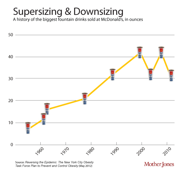
One of the contributors to the discepancy between calories in and out is that portion sizes have gone up since the 1950’s. This soft-drink diagram is taken from Mother Jones.

A genetic make-up that predisposes many people towards energy efficiency and towards overconsumption during ‘fat times’ (thrifty gene hypothesis), combined with the current abundance of calory-dense (high fat & sugar) food, the proliferation of life-styles that do not require sufficient exercise and other environmental issues can cause metabolic syndrome. Metabolic syndrome is characterized by at least 3 of the following conditions: obesity, high blood sugar, high chol. (LDL), high blood lipids (TG), and/or high blood pressure. A range of metabolic diseases are likely to follow metabolic syndrome, such as type 2 diabetes, cardiovascular disease (CVD), and many types of cancer (e.g. prostate, colon, breast).

A flow-chart summary of metabolic processes

Nutrition Metabolic Summary” by Wikimedia Commons user Boumphreyfr – Own work. Licensed under CC BY-SA 3.0.

Metabolic processes can be divided in anabolic (synthesis) and catabolic (break-down) processes. Depending on the availabel nutrient supply, the body can be said to be in an anabolic state (of energy storage), during which sugar is burnt or a catabolic state (of energy utilization), during which fat and/or amino acids are burnt.

During feeding, first, bile acid (BA) production goes up and the gall bladder empties. Then, the production of leptin and CCK signal to the brain that it should (start to) feel satieted. Meanwhile, the rise of glucose levels causes the release of insulin, which signals to cells throughout the body to take up the glucose. Some excess glucose is converted to glycogen (glucogenesis), the rest stored in adipose tissue as triglycerides (lipogenesis). When overfed, adipogenesis, takes place. Adipogenesis, the differentiation of pre-adipocytes into mature fat cells, is the means by which new adipose tissue is created.

Postprandially, during fasting, ghrelin production increases appetite. BA production goes down as the gallbladder fills. When glucose levels fall too much, glucogenolysis breaks down glucogen into glucose. After this glucose source runs out, amino acids are converted into glucose (gluconeogenesis). Most tissues can also derive their energy from fatty acids, which become available during fasting by the lysis of triglycerides (lipolysis). The brain and red blood cells, however, can only be fuelled by glucose.

Prolonged fasting (starvation) will trigger ketogenesis, the production of ketone bodies from proteins to supplement the waning free amino acid pool. Eventually, torpor (hibernation) will set in.

Nuclear receptors

Ligand-activated transcription factors are nuclear receptors that, when they bind to ligands (steroid hormones in this lecture), enhance or inhibit the transcription of certain genes. All these receptors are very similar. The family is larger than just those that bind to steroid hormones (e.g.: similar receptors have been found to bind to vitamins), such that 13% of FDA approved drugs target NRs. (For unknown reasons, C. elegans has more nuclear receptors than H. sapiens. Plants have none.)

Ligand-mediated activation: NR binds to HRE (Hormone Receptor Element), a stretch of DNA to which also an RXR receptor is bound. This complex is bound by SMRT/NCOR complex, actively repressing the HRE. When a ligand binds, it displaces the SMRT/NCOR (HDAC co-repressor) complex and the receptor complex (with the ligand) can bind to another protein complex (HAT co-activator complex), which causes acetylization of a DNA region adjacent to the HRE. (HDAC = Histone Deacetylase; HAT = Histone Acetyl Transferase.) Acetylization activates the genes.

NR example: enterohepatic circulation of bile acids

FXR (farnesoid X receptor) is also known as the bile acid receptor. FXR is expressed in the liver, which releases bile salts. Bile, produced by hepatocytes (liver cells) ends up in bile ducts, aided by 3 ABC transporters of relevance: ABCG5/8 transporters (for cholesterol), MDR2 transporters (for PC), and BSEP transporters (for BA). Bile is made up of 4% cholesterol, 24% PC and 72% BA. PC helps to neutralize BA so that it doesn’t damage cell membranes.

BA reenters the cell with the aid of a transporter called NTCP. Cholesterol also enters the hepatocytes (liver cells) (LDL through LDLR, HDL through SR-B1).

FXR activation triggers the release of bile from the hepatocyte. In enterocytes (in the illeum, the final section of the small intestine), FXR downregulates the uptake of BA by ASBT transporters and stimulates the release of BA by OSTa/b. FXR is also responsible for the release of bile salts from the illeum into the capillaries. Bile salts are also released into the blood stream by the gallbladder, which constricts under the influence of CCK.

LXR and FXR

FXR (farnesoid X receptor) is activated by bile acids, whereas LXR (liver X receptor) is activated by cholesterol.

In the liver, LXR activates ABCG5/8, inceasing the release of Chol+BA by hepatocytes. This, in turn, lowers cholesterol, as well as atherosclerosis, but also raises triglyceride production. LXR is thus a promising target for treating atherosclerosis, with the unintended side-effect of weight-gain. Indeed, a synthetic LXR ligand (GW3965) has been found to inhibit the development of atherosclerosis in LDLR and apoE knockout mice by S.B. Joseph et al. (PNAS 2002;99:7604-7609). And, indeed, the treated mice gained weight as well.

FXR activates MDR2 and BSEP transporters (which facilitate the release of PC and BA, respectively). FXR lowers cholestasis, the incidence of chol. gallstones, and the production of triglycerides; as a target for weight-loss, the unintended side-effect will be atherosclerosis.

The role of LXR & FXR in the regulation of hepatic lipid metabolism

Triglyceride (TG) from the diet or adipose tissue is hydrolysed (lipolysis) into free fatty acids (FFA), which enters the hepatocyte through the CD36 transporter. FFA in the hepatocyte can be converted into TG and stored in a complex with cholesterol, or TG+chol. can leave the hepacotye through MTTP as VLDL. Alternatively, FFAs can be used in the FAO pathway for the energy necessary to produce ketone bodies during the production of Acetyl CoA.

LXR activates lipogenesis (FFA synthesis from sugars) and increases FFA uptake from the blood, causing more TG production, which is dependent on [FFA]. ABCG5/8 transporters are also activated, increasing the release of Cholestorol and BA into the bile ducts. FXR, however, inhibits LXR and also directly inhibits lipogenesis.

PPAR (fatty acid) receptors as drug targets

Besides the FXR and the LXR receptors, to which, respectively, bile acids and oxysterols bind as ligands, Hans also treated the PPAR (Peroxisome Proliferator-Activated Receptor) family of receptors, which bind to fatty acids.

PPARγ is implicated in energy storage. PPARδ and PPARα are implicated in energy burning.

PPARα has been a drug target since the 1950s. It is activated during fasting (by PUFAs or fibrates), when it causes the burning of FFAs (through the FAO pathway) and inhibits lipogenesis. HDL goes up, while TG and FFA go down. Kersten et al. (in The Journal of Clinical Investigation 1999; 103(11):1489-1498) describe an experiment with PPARα knockout mice, which develop steatosis and hypoglycemia.

PPARγ (also an important drug target), is activated by lipids in the diet, stimulating the “energy storage” gene program, causing adipogenesis, lipogenesis, and adipokine secretion (adiponectin). When activated, PPARγ lowers blood glucose and sensitizes to insulin, making Avandia one of the top diabetes drugs in 2006. Bad press, that users might potentially suffer an increased risk of heart attacks, has made these drugs less popular.

PPARδ function remained unknown for a long time. It didn’t help that knockout mice died while still in the embryoic phase. The solution was to construct mice (VP16-PPARδ mice) in which PPARδ is only locally overexpressed, in muscle. VP16-PPARδ red muscle mass increased relative to controls.

The difference between red and white ‘meat’

Type I fibers (slow twitch), oxidation (fats), slow fatigue, long distance, “red meat”
Type II fibers (fast twitch), glycolytic (sugar), rapid fatigue, sprint, “white meat”

Transgenic mice with increased PPARδ in their muscles were true “marathon mice”. These mice are also overweight-resistent, which makes PPARδ a promesing anti-obesity drug-target. It as already used as doping by cyclists.

[See Slide 29 & 50 for a nice overview of the exam material from this lecture. Slide 29 would be nice to include here and Slide 50 would be nice to work into the introducary part of this post.]

The lecture ended with a nice citation of Mark Twain: “The only way to keep your health is to eat what you don’t want, drink what you don’t like and do what you’d rather not.”